Detection method of amyloid protein
A technology for amyloid and samples, applied in measuring devices, instruments, fluorescence/phosphorescence, etc., can solve the problems of interference in the detection process and single detection method, and achieve the effect of good biocompatibility and low cost
- Summary
- Abstract
- Description
- Claims
- Application Information
AI Technical Summary
Problems solved by technology
Method used
Image
Examples
Embodiment 1
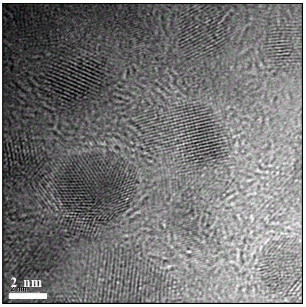
[0066] Embodiment 1: Preparation of carbon-based quantum dots
[0067] 1) Mix 1g of graphite powder with 200mL of concentrated sulfuric acid and 40g of sodium nitrate, and cool to zero. Subsequently, 4 g of potassium permanganate was slowly added, the temperature was kept at 10° C., and the mixture was vigorously stirred.
[0068] 2) After the above mixture was stirred evenly, it was transferred to an oil bath at 30°C and reacted for 50 minutes. Immediately afterwards, the temperature was raised to 140° C., and the reaction was continued for 20 h.
[0069] 3) After the reaction, the product was cooled to room temperature, diluted with 500 mL of deionized water, and adjusted to about pH 3 with Na2CO3. After filtering through a 0.22 μm membrane filter, take the supernatant. Place in a centrifuge and centrifuge at 10,000 rpm for 10 minutes. Finally, the final quantum dots were obtained by dialysis for 3 days with a dialysis bag with a molecular weight cut-off of 1000 Da.
[...
Embodiment 2
[0072] Example 2: Detection of β-amyloid monomers and fibers by carbon-based quantum dots
[0073] 1) Detection of β-amyloid monomer by carbon-based quantum dots
[0074] Aβ powder was formulated into 1 mg / mL hexafluoroisopropanol (HFIP) solution, incubated at 37°C for 3 hours, blown dry with nitrogen, and dispersed in Six Aβmonomer solutions with concentrations of 50mg / mL, 100mg / mL, 150mg / mL, 200mg / mL, 250mg / mL and 300mg / mL were formed in ultrapure water. Take 100 microliters of each of the above six solutions and mix them with 100 microliters of 1 mg / mL carbon-based quantum dot solution in a 96-well plate. Afterwards, place in a fluorescence spectrometer to measure the fluorescence intensity of the carbon-based quantum dots, set the excitation wavelength to 400nm, and the emission wavelength to 500nm. The curve of the fluorescence intensity of the carbon-based quantum dots changing with the concentration of the Aβ monomer was obtained. curve like Figure 4 As shown in (a)...
Embodiment 3
[0079] Example 3: Detection of carbon-based quantum dots on the content of β-amyloid fibers
[0080] The preparation method of Aβ monomer solution and Aβ fiber solution: same as Example 2.
[0081] 1) Mixing the prepared Aβ monomer and fiber solution according to the mass percentage of Aβ fiber from 0 to 100% to obtain Aβ monomer / fiber mixed solution with different proportions.
[0082] 2) Mix 100 microliters of the above solution with 100 microliters of carbon-based quantum dots (0.8mg / mL), add them into a 96-well plate, set the excitation wavelength to 400nm, and the emission wavelength to 450nm. The fluorescence intensity of carbon-based quantum dots in different proportions of Aβ monomer / fiber mixture was measured. The result is as Figure 5 shown. The results show that the fluorescence intensity of carbon-based quantum dots increases linearly with the increase of fiber content in the mixture.
PUM
| Property | Measurement | Unit |
|---|---|---|
| wavelength | aaaaa | aaaaa |
| wavelength | aaaaa | aaaaa |
Abstract
Description
Claims
Application Information
 - R&D Engineer
- R&D Manager
- IP Professional
- Industry Leading Data Capabilities
- Powerful AI technology
- Patent DNA Extraction
Browse by: Latest US Patents, China's latest patents, Technical Efficacy Thesaurus, Application Domain, Technology Topic, Popular Technical Reports.
© 2024 PatSnap. All rights reserved.Legal|Privacy policy|Modern Slavery Act Transparency Statement|Sitemap|About US| Contact US: help@patsnap.com


