
A microfluidic coagulation detection device and detection method thereof

A detection device and microfluidic technology, applied in biological testing, material inspection products, etc., can solve problems such as interference with optical detection, and achieve the effect of ensuring quantitative effects

Active Publication Date: 2017-05-17
常德普施康生物科技有限公司
View PDF9 Cites 0 Cited by
  • Summary
  • Abstract
  • Description
  • Claims
  • Application Information

AI Technical Summary

Problems solved by technology

[0003] The traditional optical coagulation test can only be carried out with plasma. The reason is that when red blood cells touch the reagent, they will rupture due to osmotic pressure, and then release hemoglobin, which will seriously interfere with optical detection.

Method used


Image

  • A microfluidic coagulation detection device and detection method thereof
  • A microfluidic coagulation detection device and detection method thereof
  • A microfluidic coagulation detection device and detection method thereof

Examples


Embodiment 1

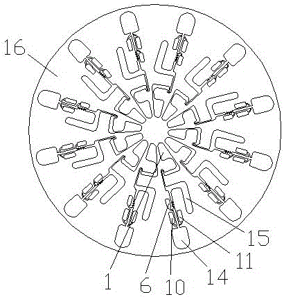
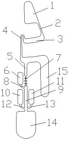
[0027] Such as figure 1 , figure 2 The microfluidic blood coagulation detection device shown includes a disc body 16 on which 12 blood coagulation detection units are installed. The blood coagulation detection unit includes a whole blood injection tank 1, and the whole blood is injected The tank 1 is connected and installed with the plasma waste tank 15, the whole blood infusion tank 1 is also connected and installed with a quick mixing unit, and the fast mixing unit is connected with the plasma waste tank 15 and installed.

[0028] The rapid mixing unit includes a plasma quantitative tank 5, which is respectively connected with the whole blood injection tank 1, the plasma waste tank 15, and the rapid mixing tank 14; wherein the rapid mixing tank 14 is respectively connected with the quality control The product injection tank 6, the reagent injection tank are connected and installed.

[0029] The plasma quantification tank 5 is connected to the rapid mixing tank 14 through the con...

Embodiment 2

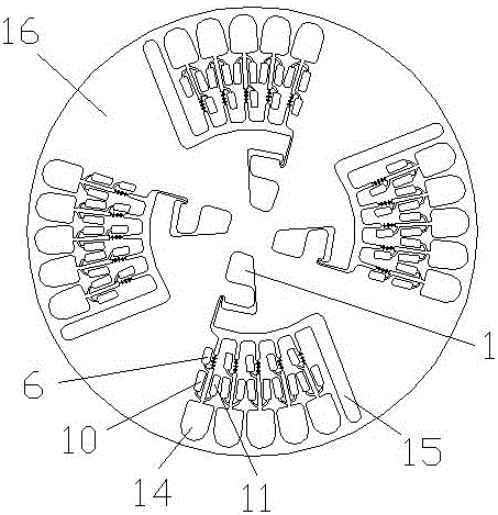
[0038] Such as image 3 , Figure 4 The microfluidic blood coagulation detection device shown includes a disk body 16 on which four coagulation detection units are mounted, and the coagulation detection unit includes a whole blood injection tank 1, the whole blood injection tank 1 is connected with the plasma waste tank 15 and installed, the whole blood infusion tank 1 is also connected with five quick mixing units, and the fast mixing unit is connected with the plasma waste tank 15 and installed.

[0039] The rapid mixing unit includes a plasma quantitative tank 5, which is respectively connected with the whole blood injection tank 1, the plasma waste tank 15, and the rapid mixing tank 14; wherein the rapid mixing tank 14 is respectively connected with the quality control The product injection tank 6, the reagent injection tank are connected and installed.

[0040] The plasma quantification tank 5 is connected to the rapid mixing tank 14 through the confluence conveying channel 9...


PUM


Abstract

The invention discloses a microfluidic blood clotting detection device and a detection method thereof. The microfluidic blood clotting detection device comprises a disc body, one or more blood clotting detection units are mounted on the disc body, each blood clotting detection unit a whole blood injection groove, the whole blood injection grooves are mounted in a mode of being communicated with blood plasma waste liquid grooves and further mounted in a mode of being communicated with one or more rapid mixing units, and the rapid mixing units are mounted in a mode of being communicated with the blood plasma waste liquid grooves. The microfluidic blood clotting detection device and the detection method thereof are high in efficiency, stable, simple, convenient and capable of conducting direct sampling on a whole blood sample, no extra sample pre-treatment is needed, and such steps as whole blood separation, quantitative blood plasma conveying and super-rapid mixing can be achieved quickly.

Description

Technical field [0001] The present invention relates to a blood coagulation detection device (optical method), in particular to a microfluidic blood coagulation detection device and a detection method thereof. Background technique [0002] Blood coagulation refers to the conversion of liquid blood into gelatinous coagulum, that is, the conversion of fibrinogen into fibrin. At present, it can be roughly divided into two categories according to the detection principle: coagulation method and substrate color method. Coagulation method: By detecting a series of physical changes in plasma under the action of a coagulation activator, such as light, mechanical movement, etc., the data obtained is analyzed by a calculator and converted into the final result, so it is also called biophysical method. Immunoturbidimetric method: Using the characteristic of specific binding between antigen and antibody, the test substance is combined with the particles labeled with its specific antibody, so...

Claims


Application Information

Patent Timeline
Patent Type & Authority Patents(China)
IPC IPC(8): G01N33/86
CPCG01N33/86
Inventor 林佳慧杨意枫余波
Owner 常德普施康生物科技有限公司
Learn More