
A high-strength bioactive porous scaffold manufacturing method

A bioactive, porous scaffold technology, applied in the fields of processing and manufacturing, medical science, prosthesis, etc., can solve the problems of difficult hole shape and size, bone can not be fully regenerated and repaired quickly, and the channel structure is not connected, so as to achieve good results. The effect of mechanical strength

Active Publication Date: 2018-11-09
杭州印生医疗科技有限公司
View PDF4 Cites 0 Cited by
  • Summary
  • Abstract
  • Description
  • Claims
  • Application Information

AI Technical Summary

Problems solved by technology

In the past, humans mainly used metals, alloys or biologically inert ceramic artificial materials with high mechanical strength to repair, fill, and replace human bone defects. Although bones have good self-regeneration capabilities, filling them has high stability and The bone at the damaged part where the biologically inert artificial implant is located cannot fully regenerate and repair itself quickly, and the filler only plays a role of mechanical support. Therefore, people began to study biodegradable, good mechanical properties, and excellent biological activity. , a material whose degradation process matches the process of human bone regeneration and repair
[0003] Recently, calcium silicate bioceramics has received more and more attention in the field of tissue engineering. Due to its excellent biological activity, porous bioceramic scaffolds have been produced by using pore-forming agent method and foam replication method, but there are It is difficult to effectively control the shape and size of the pores of the calcium silicate ceramic stent manufactured by the method, which is generally round and uneven in size, and the pore structure is not connected, the nutrients cannot be transported into the stent, and the internal metabolites cannot be smoothly Exhausted through the pores, the bone tissue cannot grow into the scaffold smoothly, and its mechanical strength is not high. With the degradation of the scaffold in the body, its strength decreases significantly. Under the stimulation of external mechanics, the structure will collapse. Unable to provide a complete stable space structure for bone tissue ingrowth, which is not conducive to bone formation in vivo

Method used


Image

  • A high-strength bioactive porous scaffold manufacturing method
  • A high-strength bioactive porous scaffold manufacturing method
  • A high-strength bioactive porous scaffold manufacturing method

Examples


Embodiment 1

[0055] Such as figure 1 Shown is a flow chart of a method for manufacturing a high-strength bioactive porous scaffold of the present invention. The specific implementation steps are as follows:

[0056] (1) Preparation of β-calcium silicate powder material and 45S5 bioglass powder material:

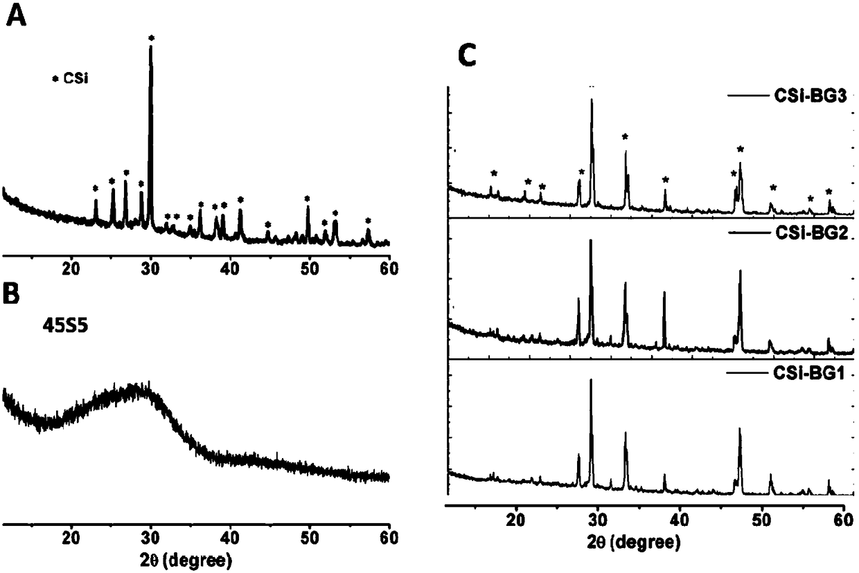
[0057] β-Calcium silicate powder material: add 1L of 0.5mol / L Ca(NO 3 ) 2 Adjust the pH value of the aqueous solution to 11.5, and then add the solution dropwise to 0.5mol / L Na with a volume of 1L 2 SiO 3 In the aqueous solution, continue to stir for 480 minutes after the addition is complete, then filter the reaction sediment, wash with deionized water 3 times, and then wash 3 times with absolute ethanol, dry at 80°C, and calcinate at 950°C for 3 hours , And then ball mill for 5 hours to obtain β-calcium silicate powder with a particle size of 1 μm to 10 μm. After X-ray diffraction test, it is proved that the powder phase is pure β-calcium silicate, such as figure 2 Shown in A.

[0058] 45S5 ...

Embodiment 2

[0072] Same as Example 1, the difference is that in step (5), the bio-ink in the extrusion unit is not vacuum defoamed, and other conditions remain unchanged. The resulting porous scaffold has many micropores on the lines, such as Figure 8 As shown, its compressive strength is 16.1 MPa, and its porosity is 65±1.5%. These micropores greatly reduce the strength of the scaffold.

Embodiment 3

[0074] Same as Example 1, the difference is that in step (11), the sintering temperature of the scaffold is changed to 1000°C for 4 hours, and other conditions remain unchanged. The compressive strength of the obtained porous scaffold is 13.2MPa, and the porosity is 68.2± 0.8%. Observed under a scanning electron microscope, its section is not dense enough, with many micropores, such as Picture 9 Shown.


PUM

PropertyMeasurementUnit
particle sizeaaaaaaaaaa
diameteraaaaaaaaaa

Abstract

The invention discloses a manufacturing method of a high-strength bioactive porous scaffold. According to the manufacturing method, process parameters in a printing process are controlled by using a three-dimensional printing device so as to extrude out bio-ink with bioactivity, ordered porous scaffolds with completely through pore passage structures are obtained by virtue of layer-upon-layer accumulation as the high-viscosity bio-ink has a self-supporting characteristic, and then the high-strength bioactive porous scaffold is obtained by virtue of a bio-glass assistant sintering process. The manufacturing method uses the three-dimensional printing device to set printing parameters randomly and control the size, shape, pore diameter and pore shape of the scaffold so as to obtain a required scaffold structure; and meanwhile, by virtue of an optimized sintering process, the scaffold still has high mechanical strength under the condition that the porosity is high. The manufacturing method disclosed by the invention is simple in operation and low in cost, and the manufactured high-strength bioactive porous scaffold can be well applied to bone tissue repair.

Description

Technical field [0001] The invention relates to the technical field of tissue engineering, in particular to a method for manufacturing a high-strength bioactive porous scaffold. Background technique [0002] The rapid and complete regeneration and repair of a series of bone injuries such as bone defects caused by external forces, bone defects caused by minimally invasive surgery, and bone tissue necrosis caused by bone tumors and inflammations are research hotspots in related fields, and are also current clinical medical problems. . In the past, humans mainly used metals, alloys or bio-inert ceramic artificial materials with high mechanical strength to repair, fill, and replace human bone defects. Although bones have good self-regeneration capabilities, these fillings have high stability and The bones of the damaged part where the biologically inert artificial implants are located cannot quickly regenerate and repair themselves completely. The fillers only play a mechanical supp...

Claims


Application Information

Patent Timeline
Patent Type & Authority Patents(China)
IPC IPC(8): B29C64/106A61L27/02A61L27/56B33Y10/00B33Y80/00
CPCA61L27/025A61L27/56B33Y10/00B33Y80/00
Inventor 贺永邵惠锋傅建中苟中入
Owner 杭州印生医疗科技有限公司
Learn More