A fluorescent-sers dual-mode super-resolution imaging probe and its preparation method and use method

A super-resolution imaging, dual-mode technology, applied in fluorescence/phosphorescence, chemical instruments and methods, material analysis by optical means, etc.

Active Publication Date: 2017-10-03
SOUTHEAST UNIV
View PDF6 Cites 2 Cited by
  • Summary
  • Abstract
  • Description
  • Claims
  • Application Information

AI Technical Summary

Problems solved by technology

However, due to the existence of the diffraction limit, the resolutions of traditional optical microscopes are only 230nm and 550nm respectively.

Method used


Image

  • A fluorescent-sers dual-mode super-resolution imaging probe and its preparation method and use method
  • A fluorescent-sers dual-mode super-resolution imaging probe and its preparation method and use method
  • A fluorescent-sers dual-mode super-resolution imaging probe and its preparation method and use method

Examples


Embodiment Construction

[0028] The present invention will be further described below in conjunction with the accompanying drawings.

[0029] The PBS buffer (Phosphate Buffer Saline, phosphate buffered saline) involved in this example has a concentration of 10 mM (the unit is millimole per liter, which can also be written as mmol / L), and pH=7.4.

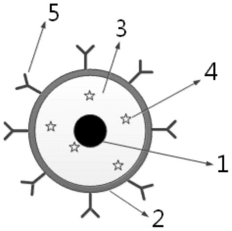
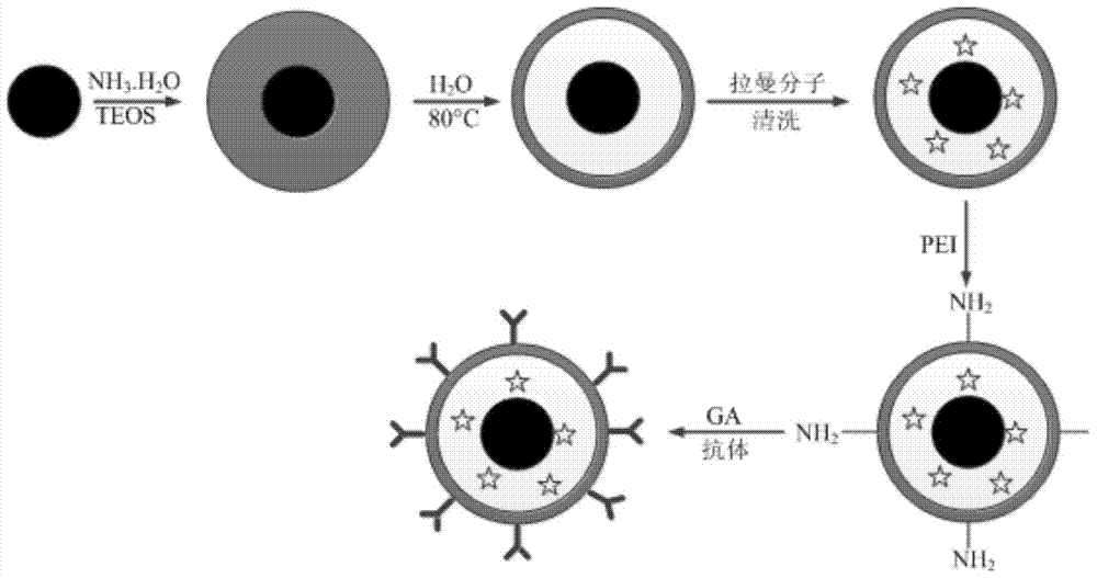
[0030] Using Nile Red (9-(diethylamino)benzo[a]phenoxazin-5(5H)-one) as the Raman molecule, the preparation process of the fluorescence-SERS dual-mode super-resolution imaging probe is as follows: figure 2 shown, including the following steps:

[0031] Step 1. Preparation of spherical gold nanoparticles

[0032] Add 200 μL of 10% chloroauric acid solution in 200 mL of deionized water, stir vigorously and heat to boiling. Then add 8 mL of 1% sodium citrate aqueous solution, and continue heating and stirring for 15 min. Stop heating, stir until the solution is cooled to room temperature, and a wine-red spherical gold nanoparticle solution is obtained.

[...


PUM


Abstract

The invention discloses a fluorescence-SERS dual-mode super-resolution imaging probe and its preparation method and use method. The fluorescence-SERS dual-mode super-resolution imaging probe is characterized in that 1, silica is coated with metal yolk-eggshell structure composite nanoparticles so that the probe is prepared, 2, a Raman molecule with a fluorescence signal is added into the cavity of the probe so that fluorescence-SERS signal switching is realized, and 3, the shell surface of the silica is labeled through a specific targeting ligand for specific organelle super-resolution optical imaging. The fluorescence-SERS dual-mode super-resolution imaging probe realizes fluorescence-SERS dual-mode super-resolution optical imaging.

Description

technical field [0001] The invention relates to a fluorescence-SERS dual-mode super-resolution imaging probe, a preparation method and a use method thereof, which belong to the super-resolution imaging technology. Background technique [0002] For a long time, far-field optical microscopy has been the most commonly used observation tool in life sciences due to its advantages of non-contact, non-damage, and ability to detect the interior of samples. However, due to the existence of the diffraction limit, the resolutions of traditional optical microscopes are only 230nm and 550nm, respectively. [0003] In order to reveal the dynamic and structural characteristics of the molecular scale in cells, improving the resolution of optical microscopy has become an urgent need for the development of life sciences. On the basis of far-field optical microscopy, scientists have invented many methods to improve imaging resolution or even exceed the resolution limit. Imaging techniques suc...

Claims


Application Information

Patent Timeline
Patent Type & Authority Patents(China)
IPC IPC(8): C09K11/02G01N21/64G01N21/65
CPCC09K11/025G01N21/64G01N21/658G01N2021/6495
Inventor 宗慎飞陆驹陈晨王著元崔一平
Owner SOUTHEAST UNIV
Why Eureka
Try Eureka