Preparation of two-color fluorescent gold bearing carbon dot and application of two-color fluorescent gold bearing carbon dot in visual inspection
A two-color fluorescence and carbon dot technology, applied in the field of applied chemistry, can solve the problems of complex and cumbersome preparation methods, and achieve the effects of obvious phenomena, fast visual detection, and high sensitivity
- Summary
- Abstract
- Description
- Claims
- Application Information
AI Technical Summary
Problems solved by technology
Method used
Image
Examples
Embodiment 1
[0029] Preparation and compositional analysis of gold-containing carbon dots. Mix 5 mL, 20 mM freshly prepared chloroauric acid solution and 1.5 mL, 100 mM glutathione solution, and dilute it to 50 mL with ultrapure water. The solution was heated while stirring in an oil bath at 80°C for 2 hours. While still hot, it was transferred to an Erlenmeyer flask containing 2 g of glucose, and heated in a household microwave oven on high for 20 minutes. Dissolve the resulting tan product in 50 mL of ultrapure water again, centrifuge (16,000 rpm, 30 min), and dialyze the supernatant (molecular weight cut-off: 8000-14000) for 24 hours to obtain a pure gold-containing carbon dot solution . Measure the UV-Vis spectrogram and fluorescence spectrogram of gold-containing carbon dots with UV-Vis spectrophotometer and fluorescence spectrophotometer, such as figure 1 , figure 2 shown; and its TEM characterization, such as image 3 As shown, it can be seen that the size of the gold-containin...
Embodiment 2
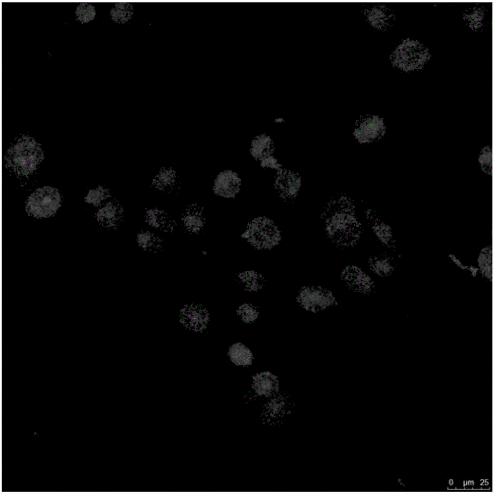
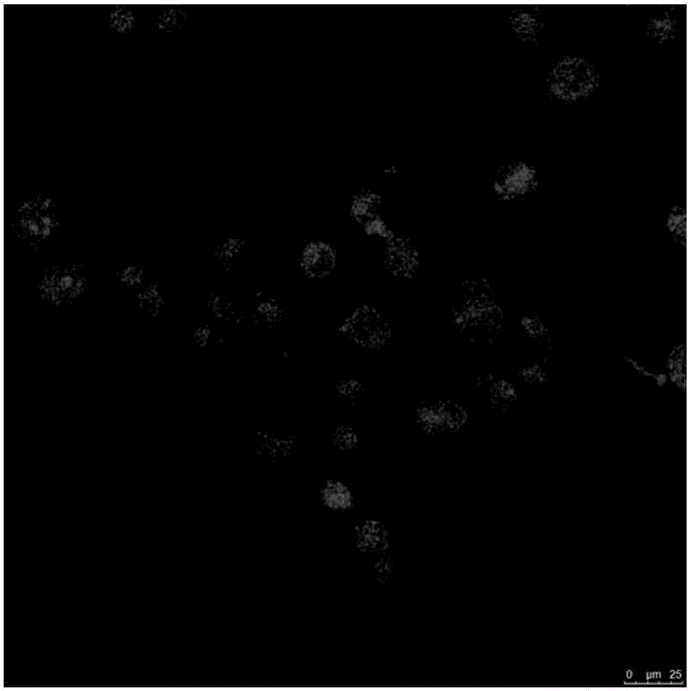
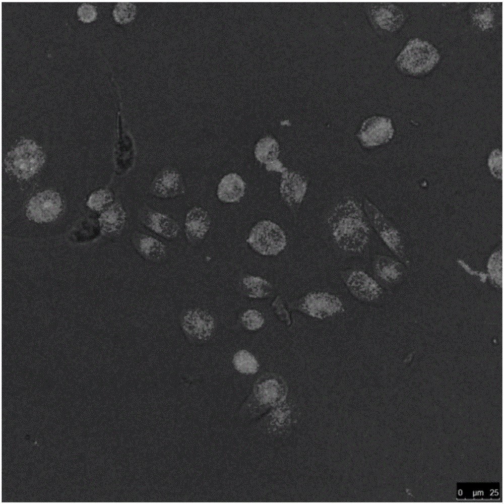
[0031] Two-color imaging of gold-containing carbon dots in cells. Dissolve gold-containing carbon dots in water to prepare a 5 mg / mL aqueous solution, then take 1 mL of gold-containing carbon dots solution and 1 mL of breast cancer phosphate buffer solution, add 9 mL of cell culture medium, and incubate at 37 ° C for 4 hours, then use Laser confocal fluorescence microscope imaging, using 405nm excitation wavelength, to obtain two-color imaging of cells, such as Figure 5 , Figure 6 , Figure 7 , Figure 8 shown.
Embodiment 3
[0033] Visual inspection of PSA: Add 30 μL, 0.1 μM single-stranded DNA aptamer with thiol-modified chain end to 3 mL of the gold-containing carbon dot solution prepared in Example 1, react for 24 hours, dialyze (molecular weight cut-off 8000~14000), to remove unreacted DNA aptamers. Since DNA is negatively charged, the zeta potential of gold-containing carbon dots is significantly reduced, as shown in Figure 9 , Figure 10 shown; Figure 11The changes in the circular dichroism spectrum shown further illustrate the successful attachment of DNA to the surface of gold-containing carbon dots. Take 10 μL of different concentrations of PSA (1, 5, 10, 50, 100, 500, 1000 ng / mL) and add it to 90 μL of the gold carbon dot solution connected with the DNA aptamer, react at 37°C for 2 h, then add 100 μL of 5 mM TMB and 100 μL, 400 μM HO 2 o 2 After reacting for 15 minutes, a color gradient change with concentration changes appears, such as Figure 12 shown; and detect its UV-Vis abs...
PUM
 Abstract
Description
Claims
Application Information
 - R&D Engineer
- R&D Manager
- IP Professional
- Industry Leading Data Capabilities
- Powerful AI technology
- Patent DNA Extraction
Browse by: Latest US Patents, China's latest patents, Technical Efficacy Thesaurus, Application Domain, Technology Topic, Popular Technical Reports.
© 2024 PatSnap. All rights reserved.Legal|Privacy policy|Modern Slavery Act Transparency Statement|Sitemap|About US| Contact US: help@patsnap.com


