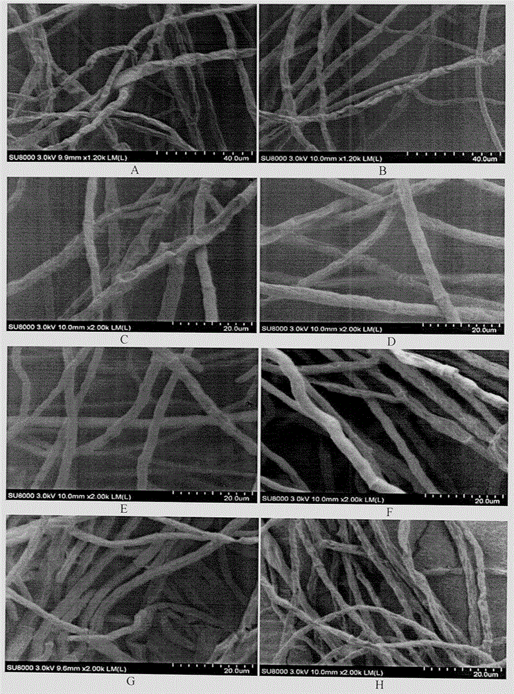
Method for preparing trichophyton scanning electron microscope sample

A technology for scanning electron microscopy and trichophyton, applied in the field of preparation of trichophyton scanning electron microscope samples, can solve problems such as insufficient contact of reagents, inaccurate judgment of results, health threats to experimenters, etc., and achieve good fixation effect

Inactive Publication Date: 2015-12-16
CHINA ACAD OF SCI NORTHWEST HIGHLAND BIOLOGY INST
View PDF13 Cites 1 Cited by
  • Summary
  • Abstract
  • Description
  • Claims
  • Application Information

AI Technical Summary

Problems solved by technology

[0003] Since the hyphae of Trichophyton are lumpy and some are thick, there is a phenomenon of insufficient contact with the reagent during the sample processing process, resulting in uneven sample processing, and the results cannot be accurately judged
Through repeated experiments, it was found that the most important step in the electron microscope sample preparation process is fixation. If the fixation effect is not good, the follow-up treatment cannot play a good role in improving it.
[0004] The most commonly used method is glutaraldehyde-osmic acid (osmium tetroxide) double immobilization at present, but osmic acid is highly toxic, and contact or inhalation can cause headache, respiratory tract inflammation, eye disease, bronchial disease, pneumonia, etc. Health poses a serious potential threat, and it is necessary to study a low-toxicity fixation method to replace the traditional double fixation method
There are also studies using glutaraldehyde alone for fixation, but it is easy to shrink the bacteria, and the effect is not satisfactory.

Method used


Image

  • Method for preparing trichophyton scanning electron microscope sample

Examples


Embodiment 1

[0018] Embodiment 1: the preparation of scanning electron microscope sample of Trichophyton mentagrophytes

[0019] 1 Materials and reagents:

[0020] 1.1 Experimental material: Trichophytonmentagrophytes

[0021] 1.2 Reagents: peptone, glucose, agar, phosphate buffer solution (PBS) at pH 7.2, 25% glutaraldehyde, ethanol, acetone, tert-butanol

[0022] 1.3 Main experimental instruments: ultra-clean bench, incubator, pipette, refrigerator, autoclave, freeze dryer, ion sputtering instrument, field emission scanning electron microscope (FESEM).

[0023] 1.4 Sample preparation

[0024] 1.4.1 Mycelia activation

[0025] SDA plate preparation: weigh 10g of peptone, 20g of glucose, and 18g of agar, dilute to 1L, sterilize at 121°C for 30min, pour the plate before the medium solidifies, and set aside;

[0026] Activation of strains: Inoculate the strains on a plate, culture in an incubator at 30°C for 5-7 days, and set aside.

[0027] 1.4.2 Sample preparation: inoculate the activ...


PUM


Abstract

The invention provides a method for preparing a trichophyton scanning electron microscope sample. When a sample is fixed, the sample is sequentially treated by at least two kinds of glutaraldehyde solution with different concentration, and the concentration of the latter glutaraldehyde is higher than that of the former glutaraldehyde. Virulent osmic acid is not used in the method, and the glutaraldehyde with gradient concentration is innovatively used for multiple times of fixing, so the fixing effect is good, the method is safe and convenient, and a new method is provided for preparing the trichophyton scanning electron microscope sample.

Description

technical field [0001] The invention relates to a preparation method of a trichophyton scanning electron microscope sample, belonging to the field of biotechnology. Background technique [0002] The pretreatment of samples is an essential process for electron microscope observation, and the quality of the treatment effect is directly related to the results of electron microscope observation. [0003] Since the hyphae of Trichophyton are lumpy and some are thick, there is a phenomenon of insufficient contact with the reagent during the sample processing process, resulting in uneven sample processing, and the results cannot be accurately judged. Through many repeated experiments, it is found that the most important step in the electron microscope sample preparation process is fixation. If the fixation effect is not good, the follow-up treatment cannot play a good role in improving it. [0004] The most commonly used method is glutaraldehyde-osmic acid (osmium tetroxide) doubl...

Claims


Application Information

Patent Timeline
IPC IPC(8): G01Q30/20
Inventor 白波曾智胡娜索有瑞徐恒
Owner CHINA ACAD OF SCI NORTHWEST HIGHLAND BIOLOGY INST
Why Eureka
Try Eureka