
A precision numerical control experimental animal image acquisition device

An image acquisition device and technology for experimental animals, applied in the field of medical research, can solve the problems of reducing reliability, bulky stereo microscopes, and difficulty in moving, etc., and achieve the effects of improving positioning accuracy, improving shooting efficiency, and quickly starting and stopping

Inactive Publication Date: 2018-09-18
FUZHOU GENERAL HOSPITAL OF NANJING MILITARY COMMAND P L A
View PDF6 Cites 0 Cited by
  • Summary
  • Abstract
  • Description
  • Claims
  • Application Information

AI Technical Summary

Problems solved by technology

[0002] In medical research, some research topics require observation and photography of experimental animal samples under a stereo microscope. Due to the high optical magnification of the stereo microscope, when shooting large-sized samples, it is necessary to take pictures in different parts, and then take pictures The process requires researchers to move large target samples, but in microscope observation, subtle movements will also be greatly magnified by the observed equipment, and it is difficult to move samples as expected during manual operation. The precise movement of the coordinates is not easy to control the time interval of the movement, which is not conducive to the microscopic observation of the researchers. When the shooting time is too long, the state of the sample may change, so that the final panoramic image will cause various problems due to the large shooting time gap. The sample status of the area does not match, which makes the picture lose its research value or reduces its credibility, and because the precision of manually moving the sample is poor, the photos taken are not accurate in area segmentation, which makes it difficult to stitch multiple photos. , if an image acquisition device that can automatically and precisely move samples can be designed, it will help to photograph experimental samples and also improve the work efficiency of researchers
[0003] Publication No. 102389291A discloses a system for collecting and analyzing experimental animal sign information and a method for collecting and analyzing it. It uses a stepping motor to move a digital camera to take pictures of samples. However, when the digital camera takes pictures of samples through a stereo microscope, due to the physical The microscope is bulky, the lens structure and observation structure are complicated, and it is not easy to move, so this method cannot be used for the observation and shooting of samples with a stereo microscope

Method used


Image

  • A precision numerical control experimental animal image acquisition device
  • A precision numerical control experimental animal image acquisition device
  • A precision numerical control experimental animal image acquisition device

Examples


Embodiment

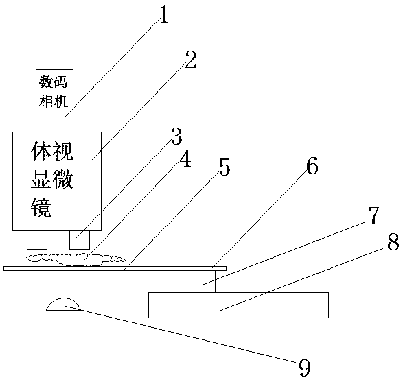
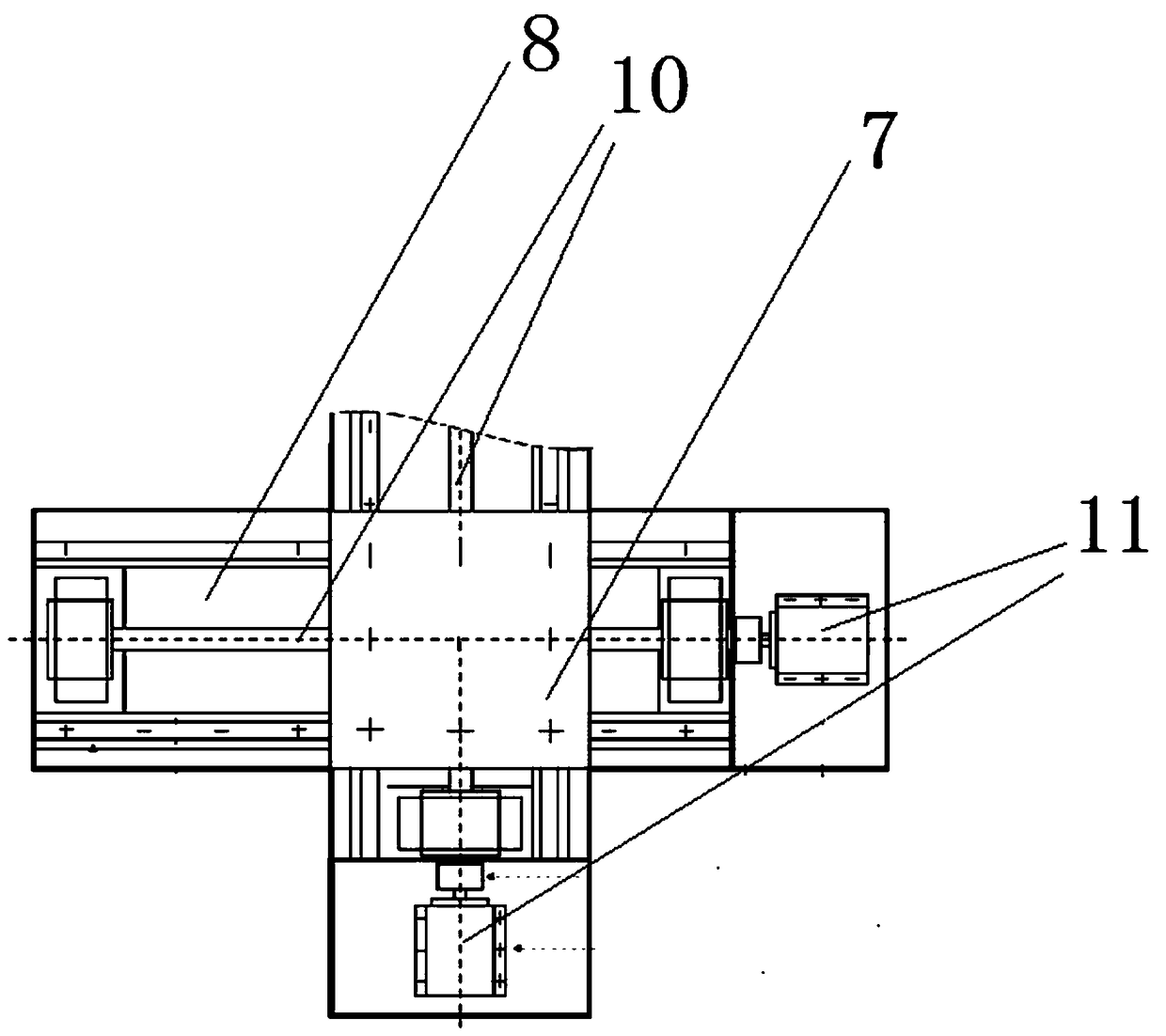
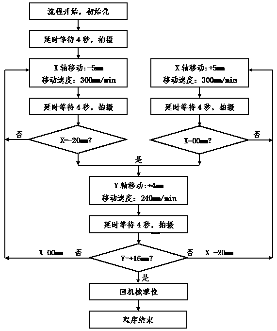
[0037] In a certain research project, it is necessary to take stereoscopic microscope images of living mouse ears under perspective light. The researcher places the anesthetized mouse on a transparent carrier plate 6, and places the mouse ear in the shooting area 5, Turn on the light source 9, adjust the light intensity of the light source 9, and adjust the stereomicroscope 2 to ensure that the mouse ear image can be clearly observed under the stereomicroscope 2, and at the same time, the light from the light source 9 can be transmitted through the mouse ear for observation To the blood vessel, adjust the sliding table system, set the bow-shaped trajectory for shooting, and make the observation image of the stereo microscope 2 be located at the starting point of the shooting trajectory, and start the shooting process, such as image 3 As shown, after the process starts and the initialization is completed, the device waits for a delay of 4 seconds to ensure that the transmission...


PUM


Abstract

The invention discloses a precise numerical control experimental animal image collecting device. The collecting device comprises a shooting module, an image processing module, a stereoscopic microscope and a slipway system. The shooting module is connected with the stereoscopic microscope, the slipway system and the image processing module. The shooting module is used for obtaining an image via the stereoscopic microscope under the control of the slipway system and transmitting the image to the image processing module. The image processing module comprises a buffer and a processor connected with the buffer. The slipway system is located at the side of the stereoscopic microscope and is driven via a driving module to slip on an X-axis direction and a Y-axis direction. A transparent carrier plate is fixedly connected on a Y-axis slipway, and one side of the transparent carrier plate adjacent to the stereoscopic microscope is stretched out of the Y-axis slipway. A shooting area for placing an experimental animal sample is located at the part of the transparent carrier plate stretched out of the slipway. The precise numerical control experimental animal image collecting device of the invention can be used for automatically performing a regional shoot to the experimental sample via the stereoscopic microscope and splicing shooting results.

Description

technical field [0001] The invention relates to the field of medical research, in particular to a precision numerical control experimental animal image acquisition device. Background technique [0002] In medical research, some research topics require observation and photography of experimental animal samples under a stereo microscope. Due to the high optical magnification of the stereo microscope, when shooting large-sized samples, it is necessary to take pictures in different parts, and then take pictures The process requires researchers to move large target samples, but in microscope observation, subtle movements will also be greatly magnified by the observed equipment, and it is difficult to move samples as expected during manual operation. The precise movement of the coordinates is not easy to control the time interval of the movement, which is not conducive to the microscopic observation of the researchers. When the shooting time is too long, the state of the sample ma...

Claims


Application Information

Patent Timeline
Patent Type & Authority Patents(China)
IPC IPC(8): H04N5/225H04N5/232G02B27/36
Inventor 郑和平吴剑威
Owner FUZHOU GENERAL HOSPITAL OF NANJING MILITARY COMMAND P L A
Learn More