
Isolated tumor fixation device and method for making pathological slices using the device

A fixed device and in vitro technology, applied in the preparation of test samples, etc., can solve problems that affect the accuracy of scanning results

Inactive Publication Date: 2017-10-31
孟雪
View PDF10 Cites 0 Cited by
  • Summary
  • Abstract
  • Description
  • Claims
  • Application Information

AI Technical Summary

Problems solved by technology

[0004] However, the existing tumor fixation devices cannot meet the above requirements, which largely affects the accuracy of scan results

Method used


Image

  • Isolated tumor fixation device and method for making pathological slices using the device
  • Isolated tumor fixation device and method for making pathological slices using the device

Examples


Embodiment 1

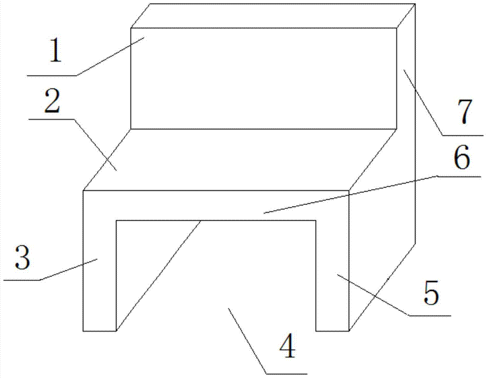
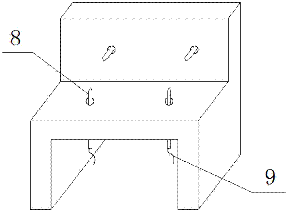
[0036] Example 1: Reference figure 1 and figure 2 As shown, an isolated tumor fixation device includes a side wall 3 and a side wall 5, and the tops of the side wall 3 and the side wall 5 are fixedly connected to the same carrying platform 6, and the side wall 3, the side wall 5 and the carrying platform 6 The rear surfaces of each are fixedly connected to a rear wall 7, a part of the rear wall 7 protrudes from the carrier platform 6, so that the front surface 1 of the part of the rear wall 7 protruding from the carrier platform 6 can fix the isolated tumor; Wherein, the upper surface 2 of the bearing platform 6 is a horizontal bearing surface for fixing the isolated tumor, and a concave cavity 4 is formed between the side walls 3 and 5 and the bearing platform 6 .

[0037] The carrying platform 6 is penetrated by vertical steel needles 8 with cotton threads 9 at the ends thereof, and the rear wall 7 is penetrated by horizontal steel needles 8 with cotton threads 9 at the en...


PUM


Abstract

The invention relates to an isolated tumor fixation device and a method for making pathological slices by using the device. The device part mainly includes: side walls located on the left and right sides; two ends of a carrying platform are respectively fixedly connected to the top surfaces of the left and right side walls; both side walls and the rear surface of the carrying platform are connected to a rear wall Fixedly connected, a part of the rear wall protrudes from the bearing platform; wherein, the upper surface of the bearing platform is a horizontal bearing surface for fixing the isolated tumor, and the front surface of the side wall is used for fixing the isolated tumor. The vertical bearing surface; a concave cavity is formed between the two side walls and the bearing platform. The device realizes the precise positioning of the isolated tumor by designing a new structure, thereby ensuring that the position of the isolated tumor in PET scanning and CT scanning is the same, and provides a guarantee for making pathological sections.

Description

technical field [0001] The invention relates to the field of medical equipment, in particular to an isolated tumor fixation device and a method for making pathological slices using the device. Background technique [0002] In isolated tumor experiments in nude mice, PET and CT scans of isolated tumors are required. [0003] Due to the limitations of current small animal PET and CT scanning equipment (PET and CT are scanned separately, and then the two sets of scanned images are fused by application software), the isolated tumor should be fixed firmly during the scanning process and ensure that the PET scan and CT scan are consistent. The placement of isolated tumors should be the same; at the same time, due to the requirements of the experiment, images obtained from PET and CT scans and pathological sections need to be compared layer by layer, which requires accurate image registration. [0004] However, existing tumor fixation devices cannot meet the above requirements, wh...

Claims


Application Information

Patent Timeline
Patent Type & Authority Patents(China)
IPC IPC(8): G01N1/28
Inventor 孟雪赵鑫张竟泽李澄明
Owner 孟雪
Learn More