Noninvasive sperm form and ultrastructure analysis method

An analysis method and ultrastructure technology, applied in the direction of material excitation analysis, Raman scattering, etc.

Inactive Publication Date: 2014-04-02
RENJI HOSPITAL AFFILIATED TO SHANGHAI JIAO TONG UNIV SCHOOL OF MEDICINE
View PDF0 Cites 11 Cited by
  • Summary
  • Abstract
  • Description
  • Claims
  • Application Information

AI Technical Summary

Problems solved by technology

At present, there is no report on the establishment of a single sperm morphology analysis method using a laser Raman spectroscopy system

Method used


Image

  • Noninvasive sperm form and ultrastructure analysis method
  • Noninvasive sperm form and ultrastructure analysis method
  • Noninvasive sperm form and ultrastructure analysis method

Examples


Embodiment

[0025] 1. Research materials and methods

[0026] 1. Experimental equipment

[0027] 1.1 Laser Raman Spectroscopy System

[0028] Dispersive confocal Raman spectrometer (Senterra R200-L), produced by Bruker, Germany, with spectral resolution ≤ 1.5cm -1 , horizontal resolution < 1um, vertical resolution < 2 um, equipped with three lasers with wavelengths of 785nm, 633nm and 532nm, three-dimensional automatic control platform and other important performances.


PUM


Abstract

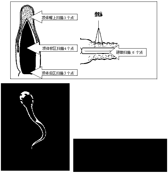
The invention relates to a noninvasive sperm form and ultrastructure analysis method. The method comprises the following steps: analyzing by using a dispersion type confocal Raman spectrometer, selecting an excitation Raman spectrum, placing sperm cells under a reflected light microscopy, and acquiring the chemical group spectrum of the sperm cells through a certain excitation power and a certain exposure time; respectively dividing the acrosome, the neck and the tail of a sperm into scanning areas, scanning three times to obtain a Raman data spectrum peak, and analyzing the waveform change and the displacement of the spectrum peak; and retrieving the acquired sperm form Raman spectrums in an RRUFF Raman database to obtain the Raman spectra of all areas of the sperm form. The laser Raman spectrum adopted in the invention is a noninvasive technology, so the characteristics of the ultrastructures of different individuals and the scattering substances of different components of each sperm can be reflected without any dye or fixation, and a new assessment method is provided for the sperm morphology analysis.

Description

technical field [0001] The invention relates to a sperm morphology and structure analysis method, in particular to a non-invasive sperm morphology and ultrastructure analysis method. Background technique [0002] At present, about 20% of couples of childbearing age in the world suffer from infertility, and male causes account for about 50% of infertility, while more than 60% of male infertility is due to oligozoospermia and asthenospermia. totally clear. At present, clinicians in many hospitals still rely on the routine semen analysis indicators in the laboratory to choose clinical treatment plans. Although the application of routine semen analysis has a long history, many medical institutions still use manual microscope analysis technology; grayscale computer-aided sperm analysis (CASA, Computer-aided sperm analysis) analysis method has been widely used clinically for many years; However, the CASA analysis method of phase contrast microscopy is still very limited in hospi...

Claims


Application Information

Patent Timeline
Patent Type & Authority Applications(China)
IPC IPC(8): G01N21/65
Inventor 李铮刘锋朱勇何琳刘宇飞
Owner RENJI HOSPITAL AFFILIATED TO SHANGHAI JIAO TONG UNIV SCHOOL OF MEDICINE
Why Eureka
Try Eureka