
Method for three-dimensional reconstruction of liver computed tomography (CT) image

A CT image and three-dimensional reconstruction technology, applied in the field of digital medical treatment, can solve the problems of poor image quality and low reconstruction efficiency

Active Publication Date: 2014-03-26
HISENSE
View PDF4 Cites 28 Cited by
  • Summary
  • Abstract
  • Description
  • Claims
  • Application Information

AI Technical Summary

Problems solved by technology

[0004] However, in related technologies, when performing three-dimensional reconstruction of liver CT images, the same reconstruction process is used for different tissue parts of the liver, resulting in low reconstruction efficiency and poor reconstructed image quality, which cannot meet the needs of doctors.

Method used


Image

  • Method for three-dimensional reconstruction of liver computed tomography (CT) image
  • Method for three-dimensional reconstruction of liver computed tomography (CT) image
  • Method for three-dimensional reconstruction of liver computed tomography (CT) image

Examples


Embodiment Construction

[0031] In order to understand the above-mentioned purpose, features and advantages of the present invention more clearly, the present invention will be further described in detail below in conjunction with the accompanying drawings and specific embodiments. It should be noted that, in the case of no conflict, the embodiments of the present application and the features in the embodiments can be combined with each other.

[0032] In the following description, many specific details are set forth in order to fully understand the present invention, but the present invention can also be implemented in other ways different from those described here, therefore, the present invention is not limited to the specific embodiments disclosed below limit.

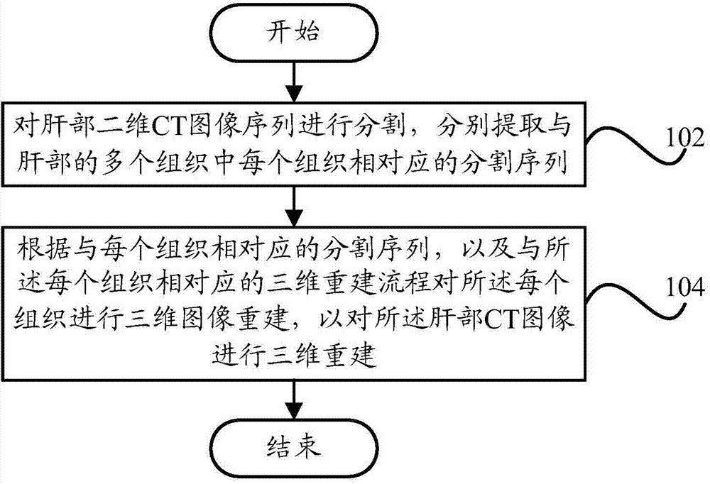
[0033] figure 1 A schematic flowchart of a method for three-dimensional reconstruction of liver CT images according to an embodiment of the present invention is shown.

[0034] Such as figure 1 As shown, the method for three-dimensional...


PUM


Abstract

The invention provides a method for three-dimensional reconstruction of a liver computed tomography (CT) image. The method comprises the steps of segmenting a liver two-dimensional CT image sequence, respectively extracting segmentation sequences corresponding to every tissue in a plurality of tissues of the liver; and performing three-dimensional image reconstruction on every tissue according to the segmentation sequences according to every tissue and three-dimensional reconstruction processes corresponding to every tissue so as to perform three-dimensional reconstruction on the liver CT image. According to the technical scheme, when the three-dimensional reconstruction is performed on the liver CT image, the corresponding reconstruction processes can be selected according to the characteristics of different tissues, and accordingly reconstruction efficiency of the liver CT image and quality of the liver CT three-dimensional reconstruction image are improved.

Description

technical field [0001] The invention relates to the field of digital medicine, in particular to a method for three-dimensional reconstruction of liver CT images. Background technique [0002] With the development of computer technology, 3D reconstruction technology based on CT (Computed Tomography, computerized tomography) images is widely used in surgical operations, such as preoperative planning, surgical simulation, and preoperative risk assessment. [0003] In traditional hepatobiliary surgery, doctors can only conduct disease diagnosis and surgical planning based on the results of two-dimensional CT scans. Diagnosis and surgical planning based on two-dimensional CT scan results are far from enough. Therefore, it is urgent to establish an intuitive and three-dimensional three-dimensional display results to guide the operation. With the help of the 3D reconstruction results of liver CT, doctors can intuitively obtain the positional relationship between hepatic veins, hep...

Claims


Application Information

Patent Timeline
IPC IPC(8): G06T17/00G06T7/00
Inventor 刘静静陈永健
Owner HISENSE
Learn More