
Method for detecting eye fundus image microaneurysm based on phase equalization

A fundus image and detection method technology, which is applied in the field of medical diagnosis, can solve problems such as uneven illumination of fundus images, difficulty in achieving detection results, and high image quality requirements, and achieve the effect of simple and practical methods, easy implementation, and reduced false detection rate

Inactive Publication Date: 2014-01-29
向日葵软件(天津)有限公司
View PDF2 Cites 20 Cited by
  • Summary
  • Abstract
  • Description
  • Claims
  • Application Information

AI Technical Summary

Problems solved by technology

Although the existing methods realize the automatic detection of MA under certain conditions, there are problems such as high false detection rate, high missed detection rate, and complex calculation.
The fundamental reason is that these methods mainly use gradient information to describe the image. Since the gradient information will change with the change of the contrast and brightness of the image, it is very sensitive to the noise in the image, so these methods have higher requirements on image quality.
Due to factors such as uneven illumination of fundus images, weak contrast, and small size of MA itself, it is difficult to exclude a large number of non-MA points by the processing method based on gradient information, and further classification based on regional feature information often increases the complexity of the system
Therefore, for fundus images with complex backgrounds, it is difficult to achieve ideal detection results based on gradient information processing methods

Method used


Image

  • Method for detecting eye fundus image microaneurysm based on phase equalization
  • Method for detecting eye fundus image microaneurysm based on phase equalization
  • Method for detecting eye fundus image microaneurysm based on phase equalization

Examples


Embodiment Construction

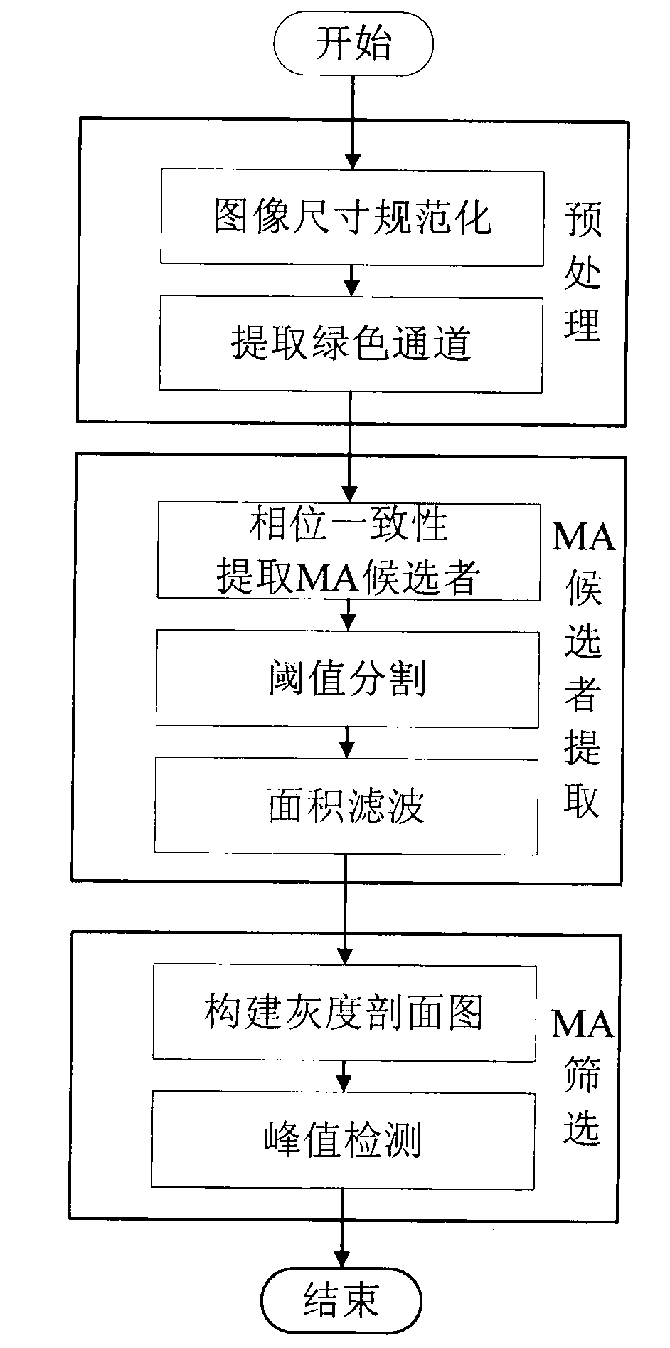
[0028] The flow chart of the present invention is as figure 1 As shown, the method first uses bilinear interpolation to normalize the image size and extracts the green channel of the color fundus image; secondly, extracts feature points based on the phase consistency model, and combines threshold segmentation and area filtering to obtain MA candidates; and finally The method of constructing a gray profile image removes irrelevant information such as blood vessels in the image and screens out the real MA. The specific implementation process of the technical solution of the present invention will be described below with reference to the accompanying drawings.

[0029] 1. Color fundus image preprocessing

[0030] 1.1 First take an original image to be detected (such as figure 2 ).

[0031] 1.2 Since the collected images may have different resolutions, in actual use, in order to preserve the image quality, bilinear interpolation is used to properly compress the original image, that is,...


PUM


Abstract

The invention relates to a method for detecting eye fundus image MA based on phase equalization. The method comprises the steps that firstly, a green channel of a colorful eye fundus image is extracted; then, feature points are extracted based on a phase equalization model, a candidate with the MA is acquired by combining a threshold segmentation and area filtering method; finally, blood vessels and other unrelated information in the image are removed through the method of constructing a gray level profile map, and therefore the true MA is screened out. The advantages of the phase equalization model are fully used, insensitivity on the brightness and the contrast ratio of the image is achieved, enhancement processing does not need to be carried out on an original image, and robustness is good; the blood vessels and other unrelated information are removed through the method of combining the gray level profile map, classification and identification are not involved, the complexity of an algorithm is greatly reduced, operating time is saved, and the MA on the colorful eye fundus image can be efficiently and automatically detected.

Description

Technical field [0001] The invention relates to a method for detecting microaneurysms in fundus images based on phase consistency, which can accurately detect fundus microaneurysms for fundus images of different image quality, belongs to the technical field of image processing and can be applied to medical diagnosis. Background technique [0002] Medical image processing and analysis have always been the focus and hot issues in the field of image processing and analysis. With the help of powerful means of graphic image technology, the quality and display method of medical images have been greatly improved, which greatly improves the level of diagnosis and treatment. Image processing technology has been introduced into ophthalmology for many years. Through the calculation and analysis of fundus images, quantitative measurement of important fundus tissues such as optic disc, retinal blood vessels, microaneurysms, and fovea can be made to make a clear distinction between normal and ...

Claims


Application Information

Patent Timeline
Patent Type & Authority Applications(China)
IPC IPC(8): G06T5/00G06K9/46
Inventor 肖志涛张芳刘璐耿磊吴骏
Owner 向日葵软件(天津)有限公司
Learn More