
Needle biopsy method based on photoacoustic imaging technique

A technology of puncture biopsy and photoacoustic imaging, which is applied in the fields of biomedicine, medical imaging and medical devices, and can solve the problems of blurred needle tip imaging, patient pain and injury, and doctor's misjudgment.

Inactive Publication Date: 2013-11-20
NANJING UNIV
View PDF6 Cites 6 Cited by
  • Summary
  • Abstract
  • Description
  • Claims
  • Application Information

AI Technical Summary

Problems solved by technology

However, the puncture biopsy technology using ultrasound imaging often causes doctors to make mistakes in judgment due to the blurred needle tip imaging, resulting in multiple punctures, which brings great pain and injury to patients.

Method used


Image

  • Needle biopsy method based on photoacoustic imaging technique
  • Needle biopsy method based on photoacoustic imaging technique
  • Needle biopsy method based on photoacoustic imaging technique

Examples


Embodiment Construction

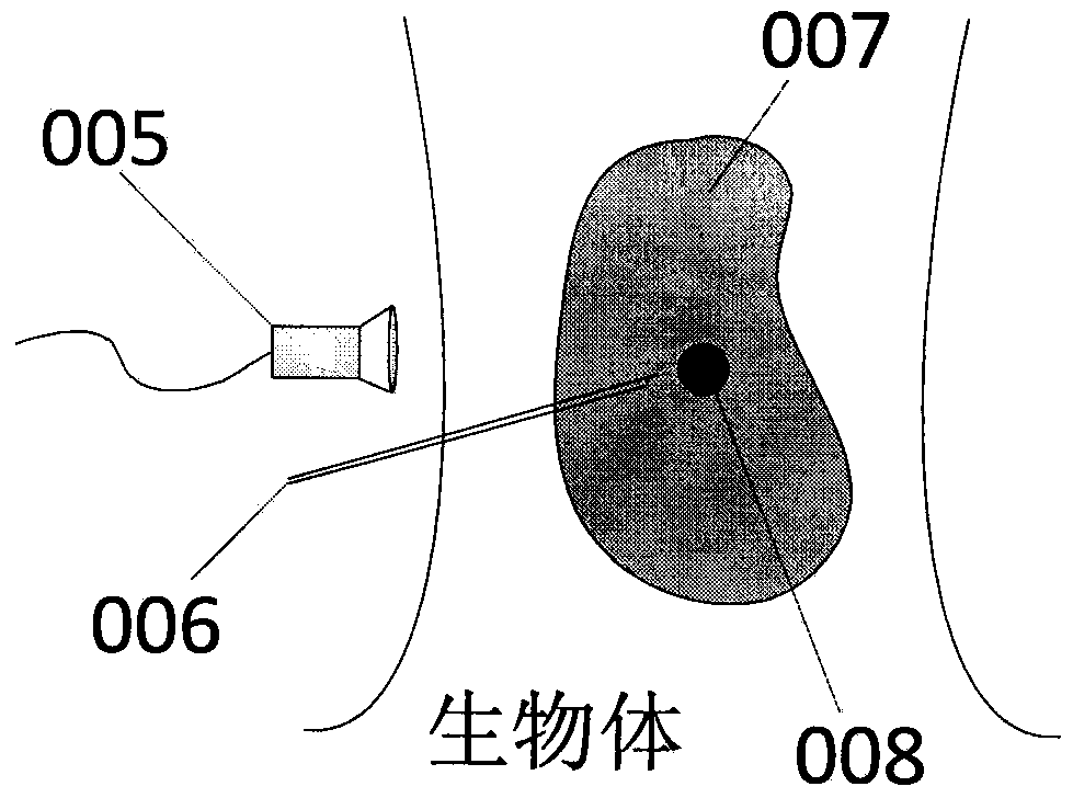
[0029] The present invention proposes a puncture biopsy method based on photoacoustic imaging technology. In this method, the image of the target tissue is obtained first by other imaging technologies, and then the laser is injected into the optical fiber embedded in the wall of the needle point for imaging, and the needle point is moved according to the imaging result.

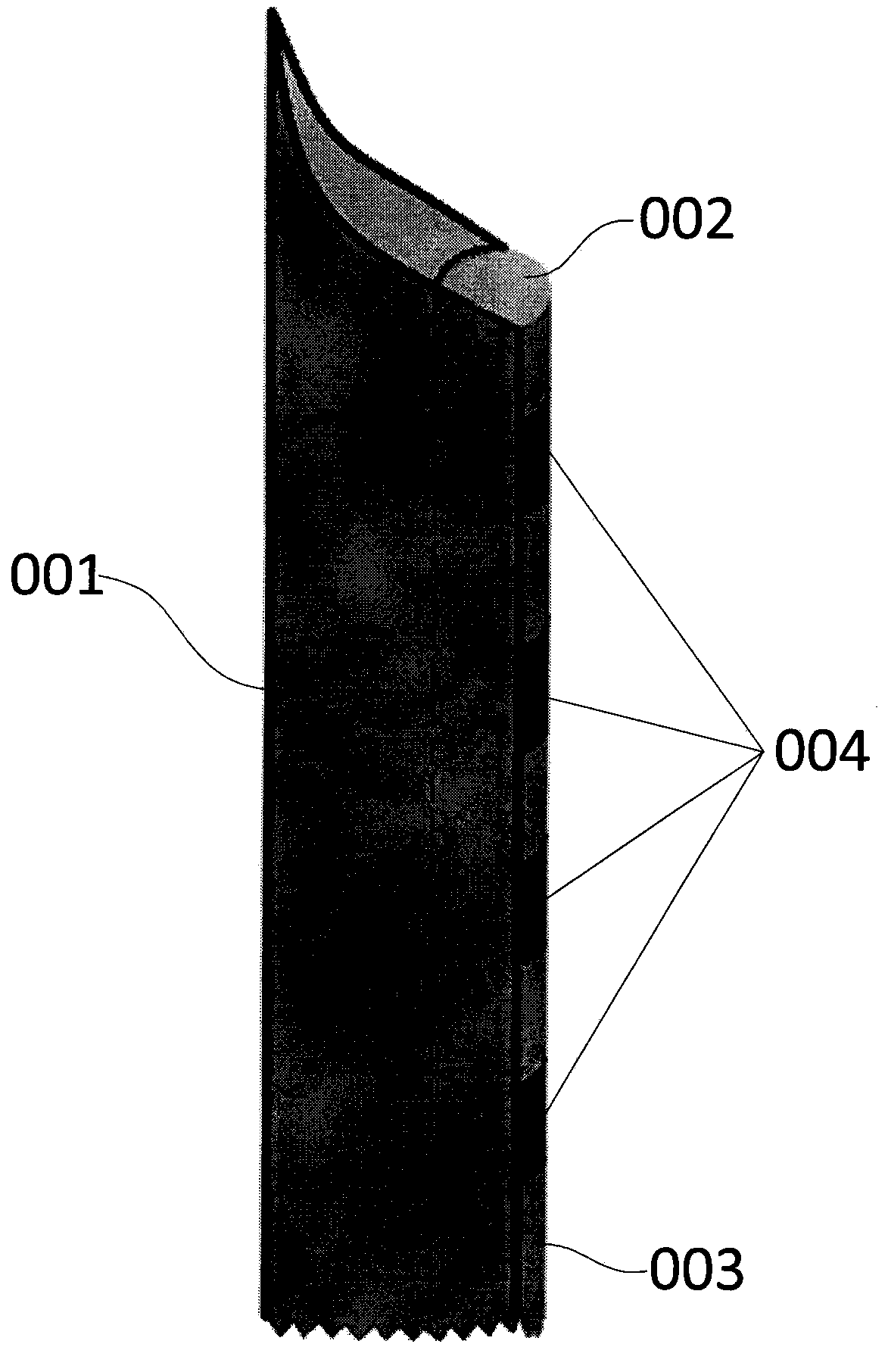
[0030] Such as figure 1 As shown, the invention discloses a puncture biopsy needle based on photoacoustic imaging technology, which includes a needle point wall (001), a laser filter membrane (002), an incident optical fiber (003), and a laser absorption layer (004).

[0031] One side of the gap in the needle point wall (001) ( figure 1 The middle right side) is slotted, the laser incident optical fiber is embedded in the slot, the needle tip is used to puncture biological tissue, and the inside of the needle tip wall can be used to obtain target tissue samples or inject drugs for treatment;

[0032] The las...


PUM


Abstract

The invention discloses a biopsy needle based on a photoacoustic imaging technique, which comprises a needle tip wall, a laser filter membrane, incident optical fibers and laser absorption layers. The invention further provides a needle biopsy method which comprises the following steps that a target tissue image is acquired in real time by other imaging techniques; the biopsy needle is aligned with a target tissue and needled; laser is emitted into the incident optical fibers; a photoacoustic signal is acquired by an ultrasonic transducer; an imaging result of a tissue nearby the laser filter membrane at the needle tip and luminous spots at the laser absorption layers and the laser filter membrane are obtained by photoacoustic reconstruction; by combining with the other imaging techniques, the target tissue imaging result and a photoacoustic imaging result of the tissue nearby the needle tip are displayed alternately; an accurate position of the needle tip is supposed by using the luminous spots at the laser absorption layers and the laser filter membrane; and when the needle tip is accurately moved to a designated position, a target tissue sample is acquired or a drug is injected for treatment. The method can accurately acquire the position of the needle tip, reduces the needling difficulty greatly, relieves a pain of a patient, and is convenient to use and outstanding in effect.

Description

technical field [0001] The invention relates to the field of biomedicine, especially the field of medical imaging and medical equipment. Background technique [0002] Since the development of ultrasound imaging, it has become one of the four modern medical imaging diagnostic technologies alongside X-CT, MRI, and PET. Compared with other medical imaging methods, it has the unique advantages of simple operation, low cost, no harm, and no trauma. It is widely used in many departments such as cardiology, obstetrics, ophthalmology, liver, kidney, gallbladder and vascular system. It has an irreplaceable role in obstetrics and gynecology. [0003] Due to its unique significance in medical diagnosis, needle biopsy technology has been widely used in pyramidal tumors and unknown tumors in various organs such as kidney, liver, lung, breast, thyroid, prostate, pancreas, testis, uterus, ovary, and body surface. biopsy, drug treatment, etc. However, the puncture biopsy technology using...

Claims


Application Information

Patent Timeline
IPC IPC(8): A61B10/02A61B5/00A61B8/00A61M5/32
Inventor 袁杰曹萌邵真天封婷张星朱毅李文超储哲琪顾鹏温馨沈庆宏都思丹
Owner NANJING UNIV
Learn More