
Organ-specific enhancement filter for robust segmentation of medical images

A medical image and filter technology, applied in the field of medical imaging, to improve health care, better segmentation accuracy, and improve reliability

Active Publication Date: 2013-04-24
KONINKLIJKE PHILIPS ELECTRONICS NV +1
View PDF5 Cites 12 Cited by
  • Summary
  • Abstract
  • Description
  • Claims
  • Application Information

AI Technical Summary

Problems solved by technology

memory storage intensity medical images

Method used


Image

  • Organ-specific enhancement filter for robust segmentation of medical images
  • Organ-specific enhancement filter for robust segmentation of medical images
  • Organ-specific enhancement filter for robust segmentation of medical images

Examples


Embodiment Construction

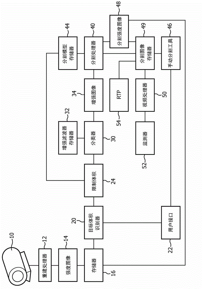
[0021] refer to figure 1 , an imaging system 10, such as a magnetic resonance imaging system, CT scanner, positron emission tomography (PET) scanner, single photon emission computed tomography (SPECT) scanner, ultrasound Scanners, radiographic imaging systems, other imaging systems, and combinations of these. The reconstruction processor 12 reconstructs the data from the imaging scanner 10 into an intensity or grayscale image 14 which is stored in a buffer or memory 16 . In a CT scan, for example, grayscale can represent the intensity or amount of radiation absorbed (or emitted) by each voxel. In a PET image, as another example, the grayscale of the intensity image can represent the intensity or amount of radioisotope events occurring in each voxel. Memory 16 may be in a workstation associated with the scanner, in a central hospital database, or the like.

[0022] The target volume identification unit 20 identifies the target volume in the intensity or grayscale image. In ...


PUM


Abstract

An imager (10) and a reconstruction processor (12) generate an intensity image (14). A region containing a target volume is identified (20) and limited (24). A classifier (30) operates on the voxels in the limited volume with an enhancement filter to generate an enhanced image, such as an image whose voxel values represent a probability that each voxel is in the target volume. A segmentation processor (40) segments the enhanced image to identify the surface of the target volume to generate a segmented image which is displayed on a monitor (52) or used in a radiation therapy planning system (54).

Description

technical field [0001] This application relates to the field of medical imaging. The present application is suitable for integration with medical image segmentation and will be described with specific reference thereto. Background technique [0002] In medical images, segmentation is the process of clearly identifying the boundaries between one or more organs or tissues through segmentation. Accurately segmenting medical images is critical for many clinical applications. However, some organs or tissues are difficult to distinguish from adjacent organs or tissues. [0003] For example, in radiation therapy, it is advantageous to deliver a relatively large dose of radiation to a target volume, such as a tumor. However, the target volume is often in close proximity and even abuts high-risk organs, which high or even moderate doses of radiation can damage. When planning the beam trajectory, it is important to know exactly the boundary between the target volume and high-risk ...

Claims


Application Information

Patent Timeline
Patent Type & Authority Applications(China)
IPC IPC(8): G06T7/00
CPCG06T2207/10081G06T2207/30004G06T7/0087G06T2207/20081G06T7/0012G06T2207/20076G06T2207/10104G06T2207/10136G06T2207/10072G06T2207/10108G06K9/34G06T7/143G06T7/149G01R33/5608G06V10/26
Inventor V·佩卡尔A·A·卡齐
Owner KONINKLIJKE PHILIPS ELECTRONICS NV
Learn More