Extraction method for metagenome
A technology of metagenomics and extraction methods, which is applied in the field of extracting metagenomics, can solve problems such as multiple times, and achieve the effects of increasing output, reducing sequencing data waste, and reducing high background interference
- Summary
- Abstract
- Description
- Claims
- Application Information
AI Technical Summary
Problems solved by technology
Method used
Image
Examples
Embodiment 1
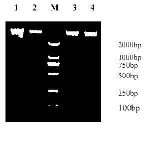
[0070] Example 1 Probe hybridization subtraction for simulated mixed samples (hybridization first and then coupling)
[0071] 1. Take 20 μl of simulated mixed DNA (according to human whole blood DNA and 2815bp plasmid mixed according to the nanogram amount of 9:1, the total amount is about 100ng / μl) in a 200 μl tube, and place it in a PCR instrument to preheat at 95°C for 5 minutes. 65°C for 5 minutes; take 27 μl of 2×hybridization solution HYB and place it in a PCR instrument to preheat at 65°C for 5 minutes;
[0072] 2. Take 0.1 μl each of the probes whose serial numbers are SEQ ID NO: 1~70, about 70 pmol, and preheat at 65°C for 2 minutes;
[0073] 3. Mix 54 μl of the above system, hybridize at 65°C for 1 hour;
[0074] 4. Take 30 μl of magnetic beads M-280 (invitrogen), wash twice with 2×B&W buffer, discard the supernatant, and resuspend with 54 μl 2×B&W;
[0075] 5. Add the hybridization system (54 μl) to the magnetic beads, mix well, and incubate at room temperature ...
Embodiment 2
[0079] Example 2 Probe hybridization subtraction for total DNA in respiratory lavage fluid samples (hybridization first and then coupling)
[0080] Except in Step 1 of Example 1, select human respiratory lavage fluid sample DNA, take 32 μl of 2×hybridization solution HYB, and take 3 μl (about 120 pmol) of each probe with sequence number SEQ ID NO: 1~4 in Step 2, Except for resuspending with 64 μl 2×B&W in step 4, the same method and conditions as in Example 1 were used to carry out first hybridization followed by coupled probe hybridization subtraction.
Embodiment 3
[0081] Example 3 Probe hybridization subtraction for simulated mixed samples (coupling first and then hybridization)
[0082] 1. Take 30μl magnetic beads M-280 (invitrogen), wash twice with 2×B&W buffer, discard the supernatant, add 15μl 2×B&W buffer to resuspend the magnetic beads;
[0083] 2. Add 0.1 μl (about 70 pmol) and 8 μl of ddH to the resuspended magnetic beads 2 O, incubate at room temperature for 15 minutes, and mix gently during the period; place on a magnetic stand at room temperature for 1 to 2 minutes, and remove the supernatant;
[0084] 3. Take 20 μl of simulated mixed DNA (the human whole blood DNA and about 2.6kbp plasmid are mixed according to the nanogram amount of 9:1, the total amount is about 100ng / μl) in a 200μl PCR tube, and put in the PCR machine to preheat at 95°C for 5 minutes , 65°C for 5 minutes; take 26 μl of 2×hybridization solution HYB and place it in a PCR instrument to preheat at 65°C for 5 minutes;
[0085] 4. Add the mixed DNA and hybr...
PUM
 Abstract
Description
Claims
Application Information
 - R&D
- Intellectual Property
- Life Sciences
- Materials
- Tech Scout
- Unparalleled Data Quality
- Higher Quality Content
- 60% Fewer Hallucinations
Browse by: Latest US Patents, China's latest patents, Technical Efficacy Thesaurus, Application Domain, Technology Topic, Popular Technical Reports.
© 2025 PatSnap. All rights reserved.Legal|Privacy policy|Modern Slavery Act Transparency Statement|Sitemap|About US| Contact US: help@patsnap.com


