
A method for pattern recognition of cancer cells using soft x-ray microscopic imaging

一种显微成像、图形识别的技术,应用在使用辐射进行材料分析、字符和模式识别、测试用样品的制备等方向

Inactive Publication Date: 2011-12-28
NO 128 HOSPITAL OF HANGZHOU
View PDF0 Cites 34 Cited by
  • Summary
  • Abstract
  • Description
  • Claims
  • Application Information

AI Technical Summary

Problems solved by technology

[0010] In China, soft X-ray microscopic imaging technology is currently mostly used in microbiology, plant and animal biological sample specimens, but there is no use of soft X-ray microscopic imaging in clinical practice and detection of cancer cells at home and abroad. Related reports on graphic recognition

Method used


Image

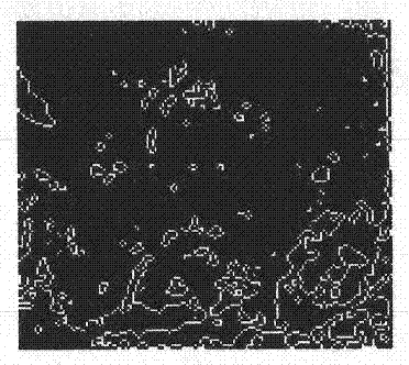
  • A method for pattern recognition of cancer cells using soft x-ray microscopic imaging
  • A method for pattern recognition of cancer cells using soft x-ray microscopic imaging
  • A method for pattern recognition of cancer cells using soft x-ray microscopic imaging

Examples


Embodiment 1

[0057] Example 1 The method for pattern recognition of esophageal squamous cell carcinoma cells using soft X-ray microscopic imaging

[0058] Selected 6 hospitalized patients with esophageal cancer (including 4 males and 2 females), aged 51-64 years (average 57.3 years).

[0059] Before the operation, fiber optic endoscopy was performed, and the pathology confirmed that it was esophageal squamous cell carcinoma, and then the operation was performed.

[0060] The resected esophageal squamous cell carcinoma specimens were sent to the pathology department for routine histological and cytological examinations. When esophageal squamous cell carcinoma cells were found, light microscopic photos of esophageal squamous cell carcinoma cells were taken under an optical microscope with a magnification of 100 times. At the same time, the specimens of esophageal squamous cell carcinoma cells were sent to the synchrotron radiation laboratory for soft X-ray microscopic beamline scanning diffr...

Embodiment 2

[0086] Example 2 The method for pattern recognition of lung cancer cells using soft X-ray microscopic imaging

[0087] Select lung cancer patients, male or female. Selected subjects need to ask about medical history, physical examination and laboratory tests such as blood routine, liver and kidney function, X-ray chest plain film, sputum to find tumor cells, serum tumor markers and soft X-ray imaging examination at the same time, And record and organize in time.

[0088] Fiberoptic bronchoscopy: Selected patients with lung tumors undergo fiberoptic bronchoscopy before surgery, extract lung tumor specimens, and send them to the pathology department for routine histological and cytological examinations. Finally, the pathological diagnosis is confirmed as lung cancer, and the pathological diagnosis results are clarified. Record and organize. The specific lung cancer specimens were taken as the research objects and sent to the National Synchrotron Radiation Laboratory of the Uni...


PUM


Abstract

This invention discloses a method for utilizing soft X-ray microimaging for cancer cell image recognition. The method comprises the steps of 1) sample preparation; 2) pathological examination; 3) soft X-ray imaging; and 4) analysis and recognition. This invention applies soft X-ray microimaging for cancer cell image recognition, successfully obtains the soft X-ray microscopic image of a cancer cell by scanning the cancer cell with synchrotron radiation soft X-ray microimaging, provides recognition steps and experimental data, and establishes a method for utilizing soft X-ray microimaging for cancer cell image recognition. This invention creates a method for analyzing soft X-ray microscopic images, provides a novel synchrotron radiation soft X-ray pathological diagnosis method for cancer diagnosis, and provides an extremely valuable basis for the creation and clinical application of soft X-ray pathology in the 21st century.

Description

technical field [0001] The invention relates to a method for pattern recognition of cancer cells by soft X-ray microscopic imaging. Background technique [0002] X-rays discovered by Roentgen in 1895 have the property of strongly penetrating matter and are suitable for imaging human tissues and organs. Its wavelength is relatively short, ranging from 0.1 to 1.0 nm. However, the interaction between X-rays and matter is very complicated, and it is difficult to manufacture optical components similar to those used in visible light applications, so it is difficult to realize X-ray microscopes. With the birth of the electron collision synchrotron radiation source, special soft X-rays are emitted with a wavelength of 1-10nm. The soft X-rays are focused and projected onto the sample through optical diffraction and optical elements, and then imaged on the image plane. [0003] The resolution of soft X-ray microscopy has reached 10nm, the principle is: [0004] The sample is placed...

Claims


Application Information

Patent Timeline
Patent Type & Authority Applications(China)
IPC IPC(8): G01N23/04G01N1/28G06K9/00G06K9/36G06N3/02
CPCG01N1/28G01N23/04G06N3/02G06K9/36G06K9/00G06V20/69
Inventor 郝建张帆董茂生罗毅吴升赵永飞王波
Owner NO 128 HOSPITAL OF HANGZHOU
Learn More