
IDL based method for three-dimensionally visualizing medical images

A medical image and three-dimensional technology, applied in the field of scientific computing visualization, can solve the problems of expensive reconstruction of local PACS systems, incompatibility of input and output interfaces of imaging equipment, and incomplete uniformity of DICOM standards, etc., and achieve friendly human-computer interaction, convenience and reliability. Scalable and efficient visualization effects

Inactive Publication Date: 2011-08-17
NANTONG UNIVERSITY
View PDF2 Cites 10 Cited by
  • Summary
  • Abstract
  • Description
  • Claims
  • Application Information

AI Technical Summary

Problems solved by technology

[0003] At present, some foreign research institutions and commercial companies have successively developed some medical image visualization analysis systems for clinical applications. Many of them depend on the hardware support provided by imaging equipment manufacturers and are expensive.
Developed countries mostly implement hardware and software double encryption processing for large-scale medical imaging equipment imported from my country, and due to many factors such as incomplete DICOM standards for various equipment, incompatibility of input and output interfaces of imaging equipment, and high cost of partial PACS system transformation, at present, my country's medical Most of the 3D visualization of images cannot be separated from the workstations of imported imaging equipment and their affiliated software platforms

Method used


Image

  • IDL based method for three-dimensionally visualizing medical images
  • IDL based method for three-dimensionally visualizing medical images
  • IDL based method for three-dimensionally visualizing medical images

Examples


Embodiment Construction

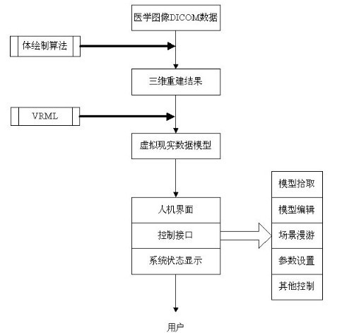
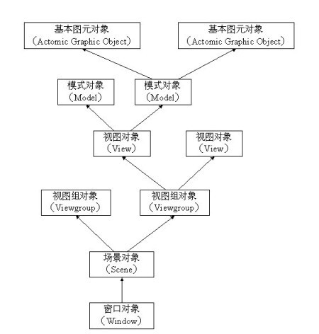
[0049] The main steps of realizing the method of the present invention please refer to figure 1 Firstly, the DICOM data of the acquired two-dimensional medical image is subjected to three-dimensional reconstruction through a volume rendering operation (algorithm) to generate a corresponding three-dimensional medical image. The volume rendering algorithm is based on IDL 6.4 object-oriented programming, which uses pre-designed and encapsulated classes provided by the IDL 6.4 language system to create various objects that meet the needs and realize corresponding control. Therefore, at the beginning of the volume rendering operation, it is necessary to define the object class in advance, that is, to define various objects in the graphics system. Each class is encapsulated in a specific visual representation, and the defined object classes include: window object IDLgrWindow, scene object IDLgrScene, view group object IDLgrViewgroup, view object IDLgrView, mode object IDLgrMode and ...


PUM


Abstract

The invention relates to an IDL based method for three-dimensionally visualizing medical images. The method comprises the following steps of: converting DICOM data into three-dimensional visualized medical images through volume rendering algorithm, converting the medical images into a virtual reality data model through VRML interactive description, and controlling the displayed graphics state through a human-computer interface on a GUI (Graphical User Interface) and a control interface so as to form visualized virtual images, wherein the volume rendering algorithm comprises the following steps of: defining an object in an object graphics system; calling the object through an object-creating OBJ_NEW function; respectively calling a pattern object, a viewing object, viewing group objects and a scene object by utilizing ADD calling format; drawing the scene object in a window object by utilizing a window object DRAW method; and completely deleting the objects by utilizing the OBJ_DESTROY function after display is accomplished. The method has the advantages of meeting the basic requirements of visualization of the medical images, and being capable of carrying out corresponding operations, such as volume rendering, slicing, and the like.

Description

technical field [0001] The invention relates to scientific computing visualization technology, in particular to a three-dimensional visualization method of medical images based on IDL language. Background technique [0002] Scientific computing visualization refers to the theory, method and technology of using computer graphics and image processing technology to convert data and calculation results in the process of scientific computing into graphics and images displayed on the screen, and interactive processing. In recent years, with the widespread use of modern medical instruments and equipment such as MRI, CT, and B-ultrasound, various medical digital images have played an increasingly important role in clinical diagnosis and medical research, and then "medical image visualization" was born. this emerging field of research. Medical image visualization technology refers to the use of artificial intelligence, pattern recognition, computer graphics, and image processing t...

Claims


Application Information

Patent Timeline
Patent Type & Authority Applications(China)
IPC IPC(8): G06T17/00
Inventor 汤敏陈峰
Owner NANTONG UNIVERSITY
Learn More