Nuclear medical imaging apparatus, image processing apparatus, and image processing method

An imaging device and image processing technology, applied in medical science, equipment for radiological diagnosis, diagnosis, etc., can solve problems such as areas that are difficult to judge objectively

Active Publication Date: 2010-11-17
TOSHIBA MEDICAL SYST CORP
View PDF9 Cites 14 Cited by
  • Summary
  • Abstract
  • Description
  • Claims
  • Application Information

AI Technical Summary

Problems solved by technology

[0011] In addition, multiple nuclear medicine images to be compared can also be displayed side by side with the difference image, but it is difficult for the doctor to judge the area of ​​interest objectively by referring to the three images for image diagnosis
[0012] In this way, the above-mentioned prior art has the problem of not necessarily assisting doctors to compare multiple examination results for image diagnosis.

Method used


Image

  • Nuclear medical imaging apparatus, image processing apparatus, and image processing method
  • Nuclear medical imaging apparatus, image processing apparatus, and image processing method
  • Nuclear medical imaging apparatus, image processing apparatus, and image processing method

Examples


Embodiment Construction

[0028] Hereinafter, preferred embodiments of the nuclear medicine imaging apparatus, image processing apparatus, and image processing method of the present invention will be described in detail with reference to the accompanying drawings. Hereinafter, a case where the present invention is applied to a PET-CT apparatus integrated with a positron emission CT apparatus (PET apparatus) as a nuclear medicine imaging apparatus and an X-ray CT (CT: Computed Tomography, computer tomography) apparatus will be described.

[0029] (Example)

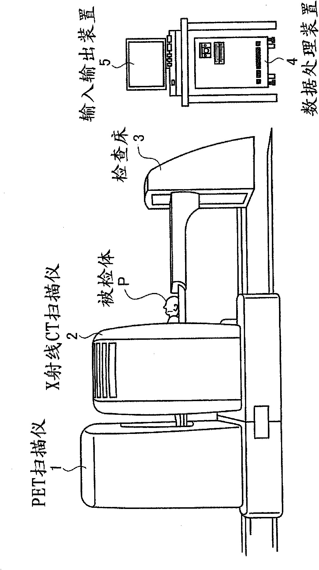
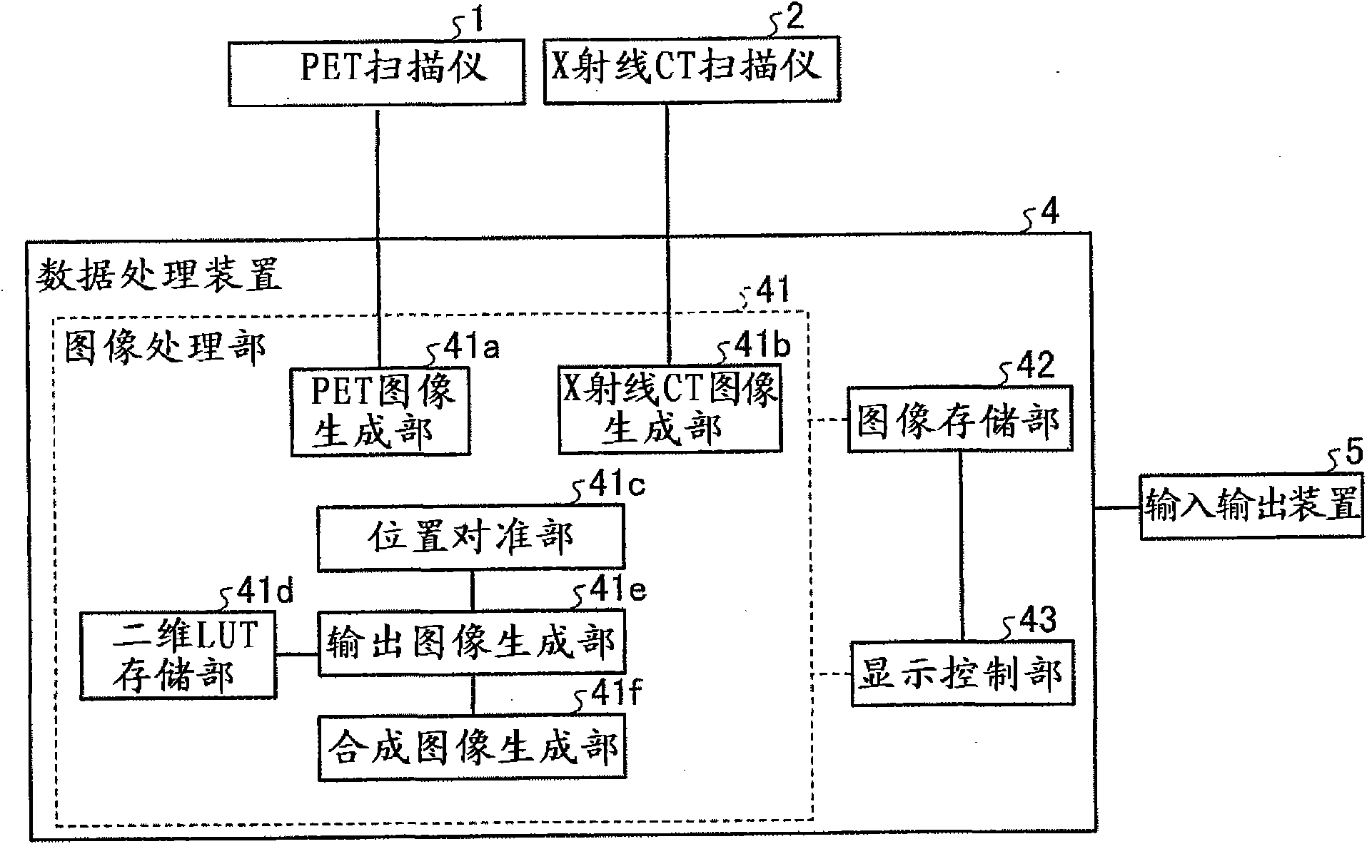
[0030] First, the structure of the PET-CT apparatus in this embodiment will be described. figure 1 It is a figure for explaining the structure of the PET-CT apparatus in this Example. Such as figure 1 As shown, the PET-CT apparatus in this embodiment is composed of a PET scanner (scanner) 1 , an X-ray CT scanner 2 , an examination bed 3 , a data processing device 4 and an input and output device 5 .

[0031] The PET scanner 1 has a ring-shaped ga...


PUM


Abstract

A two-dimensional look-up-table (LUT) storage unit stores a two-dimensional LUT in which each combination of two input values is associated with an output value for outputting a different color tone. A registration unit registers Positron Emission Tomography (PET) images of examination 1 and examination 2 by using X-ray CT images of the examination 1 and the examination 2; and an output-image creating unit acquires from the two-dimensional LUT an output value where input values are a combination of pixel values of corresponding pixels between the registered PET images, and creates an output image. Moreover, the output-image creating unit creates the output image from output values within a setting range specified on the two-dimensional LUT, among acquired output values. A display control unit controls display such that an output image or a composite image of an output image and an X-ray CT image created by a composite-image creating unit is to be displayed on a monitor.

Description

technical field [0001] This application claims the right of priority of Japanese Patent Application No. 2009-116864 filed on May 13, 2009 and Japanese Patent Application No. 2010-063222 filed on March 18, 2010, and its Japanese patent application is cited in this application all content. [0002] The present invention relates to a nuclear medicine imaging (imaging) device, an image processing device and an image processing method. Background technique [0003] Conventionally, gamma cameras (gamma cameras), Single Photon Emission (Single Photon Emission) CT (Computed Tomography, computer Nuclear medicine imaging devices such as tomography (SPECT device), positron emission (Positron Emission) CT device (PET device) and the like (for example, refer to Japanese Patent Application Laid-Open No. 2007-107995). [0004] Here, a nuclear medicine imaging device measures gamma (gamma) rays emitted from isotopes or labeled compounds selectively introduced into living tissue from outsi...

Claims


Application Information

Patent Timeline
Patent Type & Authority Applications(China)
IPC IPC(8): A61B6/03
CPCA61B6/5235A61B6/037A61B6/5247A61B6/032
Inventor 野边昌史关将俊
Owner TOSHIBA MEDICAL SYST CORP
Why Eureka
Try Eureka