
Ultrasonic imaging method and device

An ultrasonic imaging method and image technology, applied in ultrasonic/acoustic/infrasonic diagnosis, acoustic diagnosis, infrasonic diagnosis, etc., can solve problems such as complex processing process, detection of echo interference, waste of pulse width and frequency bandwidth resources, and achieve mutual Good correlation, elimination of interference, and energy-enhancing effects

Inactive Publication Date: 2010-10-27
SHENZHEN UNIV
View PDF4 Cites 49 Cited by
  • Summary
  • Abstract
  • Description
  • Claims
  • Application Information

AI Technical Summary

Problems solved by technology

[0009] However, in practical applications, the above-mentioned method of generating acoustic radiation force has disadvantages: the excitation pulse that generates acoustic radiation force will cause great interference to the detection echo after reflection
[0011] Although the above two solutions can solve the interference problem, the processing process is relatively complicated, which wastes pulse width and frequency bandwidth resources.

Method used


Image

  • Ultrasonic imaging method and device
  • Ultrasonic imaging method and device
  • Ultrasonic imaging method and device

Examples


Embodiment Construction

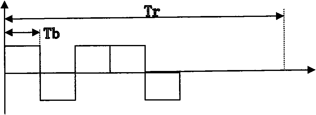
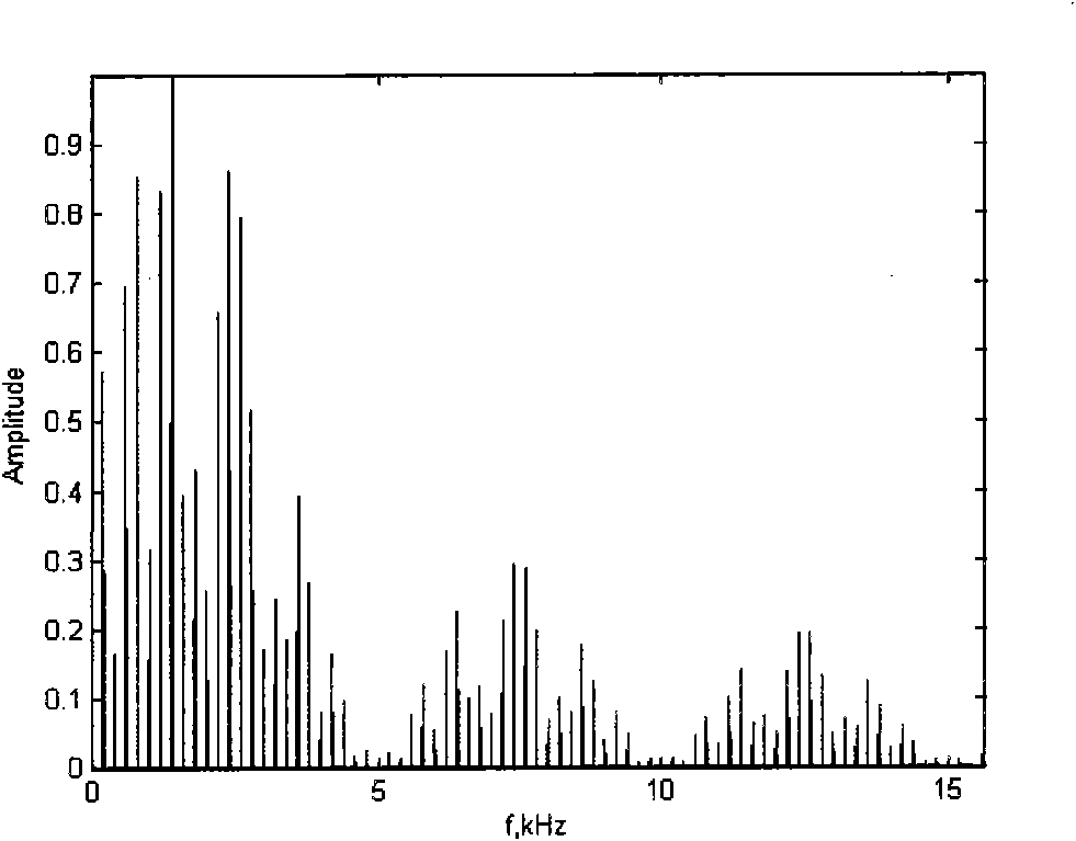
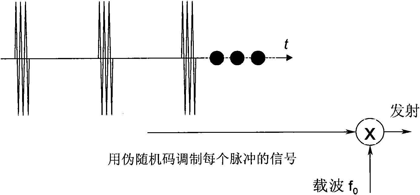
[0054] The present invention provides an ultrasonic imaging method and device that uses coded excitation pulses to generate acoustic radiation force. In order to make the purpose, technical solutions and effects of the present invention clearer and clearer, the present invention will be further described in detail below with reference to the accompanying drawings and examples. It should be understood that the specific embodiments described here are only used to explain the present invention, not to limit the present invention.

[0055] In medical ultrasound, the frequency response of biological soft tissue is mainly in the low frequency part, and the amplitude of the vibration displacement generated by the response is very small (micron level). The vibration amplitude is determined by the amplitude of the acoustic radiation force. Therefore, in order to improve the accuracy of detection, the acoustic radiation force should have more low-frequency components, smaller frequency ...


PUM


Abstract

The invention discloses ultrasonic imaging method and device. The method comprises the following steps of: firstly carrying out coded modulation on a pulse generating an acoustic radiation force, and exciting a probe to emit an ultrasonic wave to generate a multi-frequency acoustic radiation force to generate tissue vibration; then adopting the coded modulation different from an exciting pulse for the pulse detecting the tissue vibration, ensuring the good correlation between two code words, and eliminating the interference caused by an exciting pulse echo generating the acoustic radiation force for a detection echo by utilizing a CDMA (Code Division Multiple Access) technology similar to communication, thereby improving the signal-to-noise ratio and increasing the energy of the acoustic radiation force by more fully utilizing the pulse width and the frequency band width resources of a system.

Description

technical field [0001] The invention relates to an ultrasonic imaging method and device applicable to the fields of ultrasonic nondestructive testing, medical ultrasound, ultrasonic elastography and the like. Background technique [0002] The basic principle of elastic measurement is to apply a pressure to the tissue. Under the action of elastic mechanics, biomechanics and other laws, the tissue will produce a response, including displacement, deformation, etc. The response of tissues with different elastic modulus or hardness will be different. Ultrasound, nuclear magnetic resonance or optical methods are used to detect the response inside the tissue, so as to reflect the elastic properties of the tissue. [0003] Acoustic radiation force elastography is a type of ultrasound elastography. Ultrasonic waves are emitted to an object, and the absorption and scattering of the incident ultrasonic waves by the object causes a change in the sound energy density, thereby generating...

Claims


Application Information

Patent Timeline
IPC IPC(8): A61B8/00G01N29/14
Inventor 覃正笛陈思平汪天富曾斯宁
Owner SHENZHEN UNIV
Learn More