Eureka AIR delivers breakthrough ideas for toughest innovation challenges, trusted by R&D personnel around the world.

Method for processing fundus images

A fundus image and image technology, applied in image data processing, ophthalmoscopy, image enhancement, etc., can solve problems such as imperfection, poor patient comfort, and high impact on inspection results, to improve quality, improve the level of diagnosis and treatment, and improve medical treatment. Diagnostic-level effects

Inactive Publication Date: 2010-06-16
中国大恒(集团)有限公司
View PDF2 Cites 12 Cited by
  • Summary
  • Abstract
  • Description
  • Claims
  • Application Information

AI Technical Summary

Problems solved by technology

The current ophthalmic diagnosis and treatment mostly use close-up examinations between doctors and patients, the comfort of patients is poor, and the examination results are highly affected by human factors
So far, the technology of fundus image matching, optic nerve measurement, and fundus image stitching based on machine vision technology is still quite imperfect in the medical field.

Method used


Image

  • Method for processing fundus images
  • Method for processing fundus images

Examples


Embodiment Construction

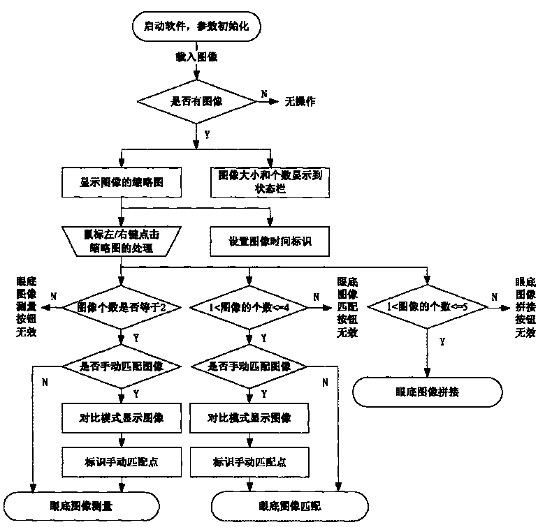
[0014] The invention provides a fundus image processing system based on machine vision, which is used for image matching of fundus images generated in ophthalmology, optic nerve measurement and fundus image splicing, and can realize real-time analysis, diagnosis, storage and transmission of fundus images of patients. The present invention will be described in detail below in conjunction with the accompanying drawings and specific embodiments. The fundus image processing method provided by the present invention can realize the following functions:

[0015] 1. Automatic matching of multiple images, including image correction, cropping the region of interest, automatic generation of multi-frame images in two different formats, jpeg and gif, and the processing time of the four images is controlled within 500ms;

[0016] 2. The optic nerve measurement of two images has the function of automatically finding the optic cup and optic disc, and assists in manual search. It can accuratel...


PUM


Abstract

The invention is a method for processing fundus images based on machine vision. The method comprises the following steps of: loading collected fundus images; opening the collected fundus images, matching the fundus images; measuring the fundus images, and splicing the fundus images, wherein the image matching comprises automatic matching and manual matching; carrying out the measurement of the fundus after the completion of matching, cutting the interested region of the matched images, setting as a comparison mode to display two images, and then manually setting a characteristic region in the images, measuring the area of the fundus characteristic region in two images; the fundus image splicing means that a plurality of fundus images are loaded in the eyes of one person to automatically splice, so that a doctor can see the full view of the fundus images of a patient. The invention can realize the functions of real-time analysis, diagnosis, storage and transmission of the fundus images of the patient, thereby assisting the diagnosis of an ophthalmologist to improve the accuracy and the convenience of the diagnosis.

Description

technical field [0001] The invention relates to designing an image processing method, in particular to a fundus image processing method for fundus image matching, optic nerve measurement and fundus image splicing. Background technique [0002] With the development of technology and the growth of the market, the hardware and software of machine vision technology are becoming more and more perfect, and the development of machine vision technology is very rapid. From the 1950s with statistical recognition and analysis of two-dimensional images as the main function, it developed to image shading, texture, motion and imaging geometry. By the 1970s, visual application systems had appeared. Later, with the rapid development of computer technology and the continuous maturity of computer vision theory, intelligent machine vision systems gradually appeared. Digital image processing is widely used in electronics, printing, medicine, food and other manufacturing industries, especially ...

Claims


Application Information

Patent Timeline
Patent Type & Authority Applications(China)
IPC IPC(8): A61B3/12G06T5/00G06T7/00
Inventor 朱江兵朱瑞慧
Owner 中国大恒(集团)有限公司
Why Eureka
Eureka Blog
Learn More