
Self-adaptive ultrasonic imaging method for inhibiting tissue flicker, and device thereof

An adaptive tissue image technology, applied in ultrasound/sound wave/infrasonic wave diagnosis, application, image enhancement, etc., can solve the problems of large tissue energy, large difference in parameter distribution, strong parameter sensitivity, etc., and achieve accurate tissue movement speed , Inhibit tissue flicker, judge reliable effect

Active Publication Date: 2009-09-09
SHENZHEN MINDRAY BIO MEDICAL ELECTRONICS CO LTD
View PDF0 Cites 16 Cited by
  • Summary
  • Abstract
  • Description
  • Claims
  • Application Information

AI Technical Summary

Problems solved by technology

Similar to the problem encountered in spectrum shifting, when the estimated tissue motion velocity cannot correctly reflect the real tissue motion, the cut-off frequency of the wall filter set only by the tissue motion frequency will be low, which also leads to the deviation of the residual tissue energy after wall filtering. large, eventually showing tissue flickering on the image
The disadvantage of dual-channel is that two different parameter distributions (energy, velocity, variance) are used to judge tissue flickering. The parameter distributions of different people and different detection parts are too different, which makes this method too sensitive to parameters.

Method used


Image

  • Self-adaptive ultrasonic imaging method for inhibiting tissue flicker, and device thereof
  • Self-adaptive ultrasonic imaging method for inhibiting tissue flicker, and device thereof
  • Self-adaptive ultrasonic imaging method for inhibiting tissue flicker, and device thereof

Examples


Embodiment Construction

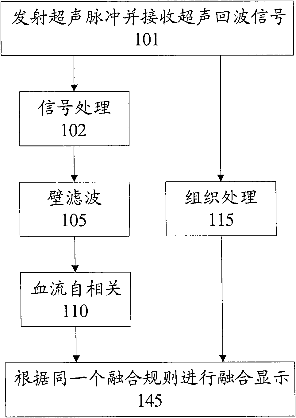
[0044] figure 1 is a flowchart of a conventional ultrasound imaging method that does not include adaptive tissue flicker suppression. In step 101, an ultrasonic pulse is transmitted to an object to be detected and an ultrasonic echo signal is received from the object to be detected. In step 102, the ultrasonic echo signal is processed to obtain an audio signal. In step 105, wall filtering is performed on the audio signal. In step 110, blood flow autocorrelation is performed on the wall-filtered audio signal to obtain the velocity, energy and variance of the blood flow. In step 115, the ultrasonic echo signal is processed to obtain tissue image data. In step 145, the velocity, energy and variance of the blood flow are fused with the tissue image data for display according to the same fusion rule, so as to provide the user with a two-dimensional image that simultaneously includes tissue structure and blood flow dynamics.

[0045] When focusing on tissue flicker caused by hum...


PUM


Abstract

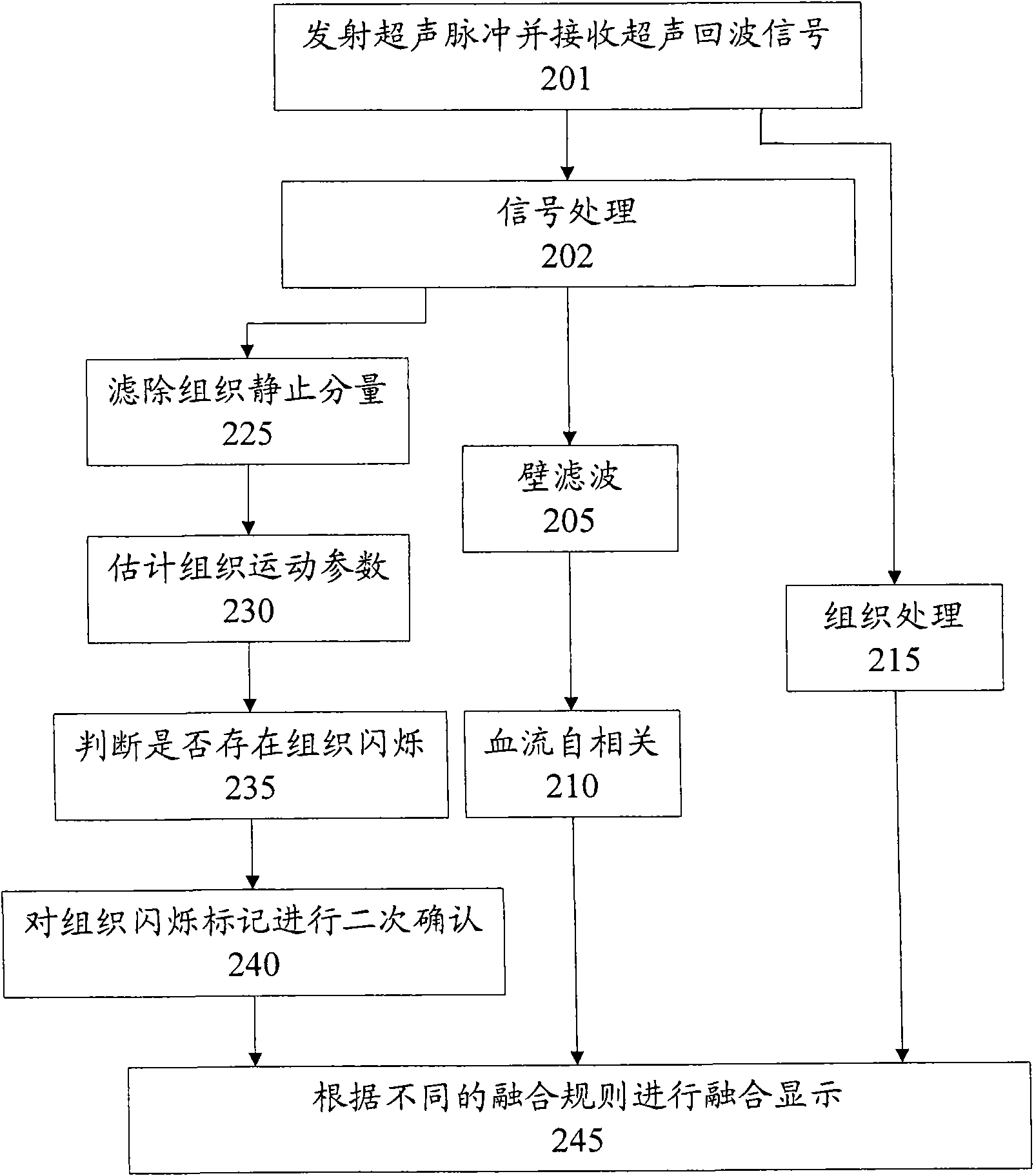
The invention provides a self-adaptive ultrasonic imaging method for inhibiting tissue flicker, and a device thereof. The method and the device transmit ultrasonic pulses to an object to be detected, receive ultrasonic echo signals from the object to be detected, process the ultrasonic echo signals so as to obtain tissue image data, process the ultrasonic echo signals so as to obtain audio signals, process the audio signals so as to obtain blood-flow image data, and display the tissue image data or the blood-flow image data according to fusion rules, wherein a first fusion rule is adopted forthe parts with tissue flicker in the object to be detected; a second fusion rule is adopted for the parts with no tissue flicker in the object to be detected; and the first fusion rule differs from the second fusion rule. The method and the device can effectively weaken and even eliminate the tissue flicker caused by various tissue movements in ultrasonic imaging systems.

Description

technical field [0001] The present invention relates to the field of ultrasound imaging, and more particularly, to an ultrasound imaging method and device for adaptively suppressing tissue flicker. Background technique [0002] The color ultrasound imaging system uses the Doppler effect to detect the movement parameters of blood flow and organs in the human body. The Doppler frequency shift of human tissue relative to the probe will be caused by the heartbeat, respiration, periodic contraction and relaxation of the blood vessel wall or the movement of the probe relative to the human body. The situation of this kind of tissue movement is relatively complicated, it can be a single tissue movement relative to the probe, it can also be a reciprocating tissue movement relative to the probe, and it can even be twisting. For a single tissue motion, when the Doppler frequency shift of the tissue motion is higher than the Doppler frequency shift of the slow blood flow to be detected...

Claims


Application Information

Patent Timeline
IPC IPC(8): A61B8/06
CPCA61B8/461A61B8/13G06T2207/20012G06T2207/10132G06T5/002G06T2207/30104G06T5/001A61B8/06G06T5/70
Inventor 董永强
Owner SHENZHEN MINDRAY BIO MEDICAL ELECTRONICS CO LTD
Learn More