Construction method of body surface fistula rat animal model
A technology of animal model and construction method, which can be applied to preparations for in vivo experiments, pharmaceutical formulations, medical science and other directions, can solve the problem of no clinical research on animal models of fistula, and achieve good stability.
- Summary
- Abstract
- Description
- Claims
- Application Information
AI Technical Summary
Problems solved by technology
Method used
Image
Examples
Embodiment 1
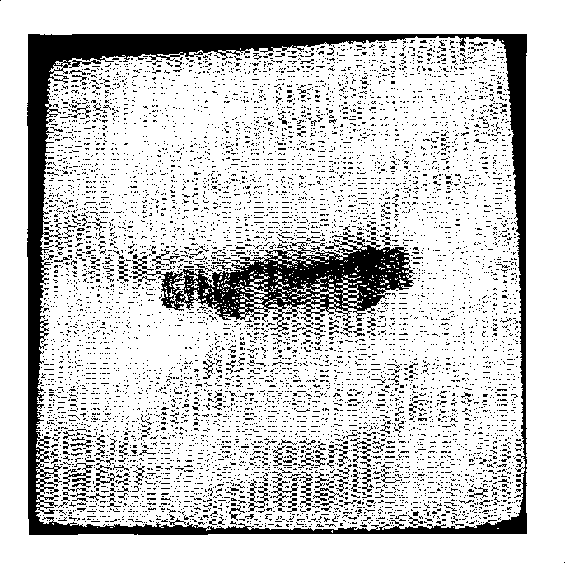
[0021] Experimental animals: 30 SD (Sprague-Dawly) rats of about 350 grams were randomly divided into three groups: Staphylococcus aureus group, Escherichia coli group, and mixed bacteria group. The incisions were made at a distance of 3 cm, and 3 cm long sterile medical gauze was embedded between them, and the two ends were sutured to the skin for fixation.
[0022] 1. Staphylococcus aureus group: inject 3×10 7 0.2ml of Staphylococcus aureus at a concentration of cfu / ml was implanted into rat gauze.
[0023] 2. Escherichia coli group: use a syringe to inject 3×10 7 0.2ml of Escherichia coli at a concentration of cfu / ml was implanted into rat gauze.
[0024] 3. Mixed bacteria group: use a syringe to inject 3×10 7 0.2ml of mixed bacteria of Staphylococcus aureus and Escherichia coli at the concentration of cfu / ml were implanted into the rat gauze.
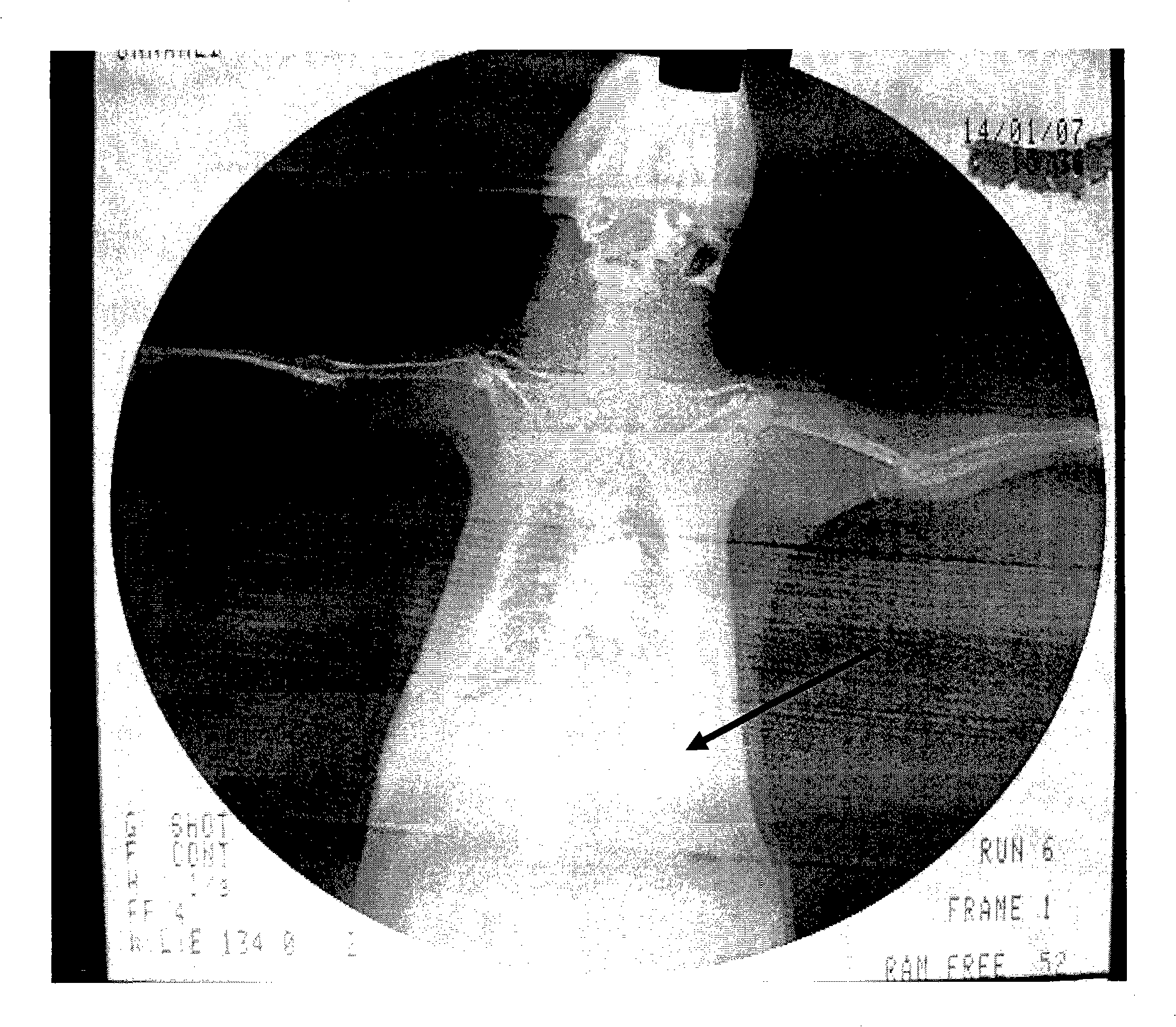
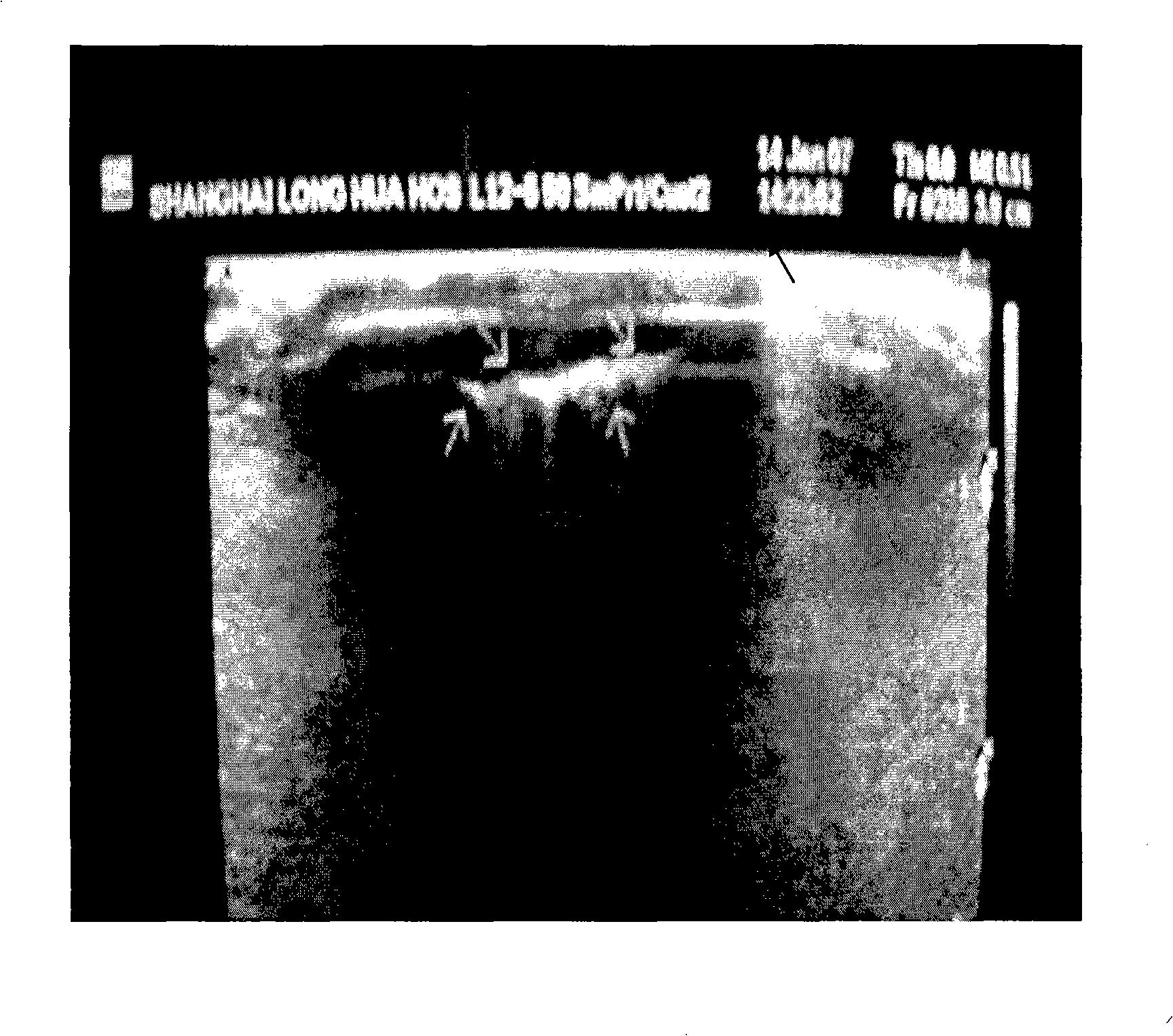
[0025] After inoculation, the rats in each group were observed daily for body temperature changes, local wound manifestations,...
Embodiment 2
[0028] Experimental animals: 30 SD (Sprague-Dawly) rats of about 350 grams were randomly divided into three groups: Staphylococcus aureus group, Escherichia coli group, and mixed bacteria group. The incisions were made at a distance of 3 cm, and a 3 cm long self-made spring gauze was embedded between them, and the two ends were sutured to the skin for fixation.
[0029] 1. Staphylococcus aureus group: inject 3×10 9 0.2ml of Staphylococcus aureus at a concentration of cfu / ml was implanted into rat spring gauze.
[0030] 2. Escherichia coli group: use a syringe to inject 3×10 9 0.2ml of Escherichia coli at a concentration of cfu / ml was implanted into rat spring gauze.
[0031] 3. Mixed bacteria group: use a syringe to inject 3×10 9 0.2ml of mixed bacteria of Staphylococcus aureus and Escherichia coli at a concentration of cfu / ml were implanted into rat spring gauze.
[0032] After inoculation, the body temperature of the rats in each group was observed daily; local wound man...
Embodiment 3
[0035] Experimental animals: 30 SD (Sprague-Dawly) rats of about 350 grams were randomly divided into three groups: Staphylococcus aureus group, Escherichia coli group, and mixed bacteria group. The incisions were made at a distance of 3 cm, and a 3 cm long self-made spring gauze was embedded between them, and the two ends were sutured to the skin for fixation.
[0036] 1. Staphylococcus aureus group: inject 9×10 8 0.2ml of Staphylococcus aureus at a concentration of cfu / ml was implanted into rat spring gauze.
[0037] 2. Escherichia coli group: inject 9×10 8 0.2ml of Escherichia coli at a concentration of cfu / ml was implanted into rat spring gauze.
[0038] 3. Mixed bacteria group: use a syringe to inject 9×10 8 0.2ml of mixed bacteria of Staphylococcus aureus and Escherichia coli at a concentration of cfu / ml were implanted into rat spring gauze.
[0039] After inoculation, the body temperature of the rats in each group was observed daily; local wound manifestations and f...
PUM
 Abstract
Description
Claims
Application Information
 - R&D
- Intellectual Property
- Life Sciences
- Materials
- Tech Scout
- Unparalleled Data Quality
- Higher Quality Content
- 60% Fewer Hallucinations
Browse by: Latest US Patents, China's latest patents, Technical Efficacy Thesaurus, Application Domain, Technology Topic, Popular Technical Reports.
© 2025 PatSnap. All rights reserved.Legal|Privacy policy|Modern Slavery Act Transparency Statement|Sitemap|About US| Contact US: help@patsnap.com


