Porcine circovirus and helicobacter combination vaccines and methods of use
A porcine circovirus and Helicobacter technology, applied in the direction of virus/bacteriophage, virus, vaccine, etc., can solve the problems of microbial flora disorder and other problems
- Summary
- Abstract
- Description
- Claims
- Application Information
AI Technical Summary
Problems solved by technology
Method used
Image
Examples
Embodiment 1
[0244] Example 1: Method for Isolating and Characterizing PCV Isolates
[0245] 1) Cell culture: PCV-free PK15 derivative, ie Dulac cell line, was obtained from Dr. John Ellis (University of Saskatchewan, Saskatoon, Saskatchewan). The Vero cell line was obtained from the American Type Culture Collection (ATCC), Manassas, VA. These cells were cultured in medium as recommended by ATCC and incubated at 37 °C and 5% CO 2 incubate.
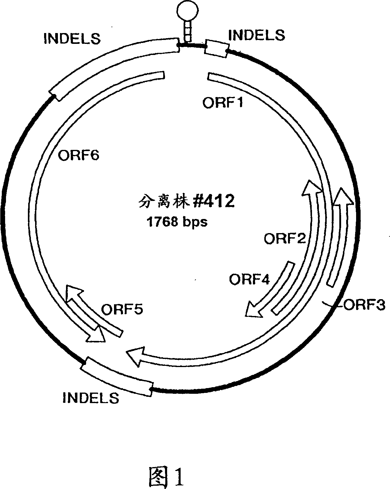
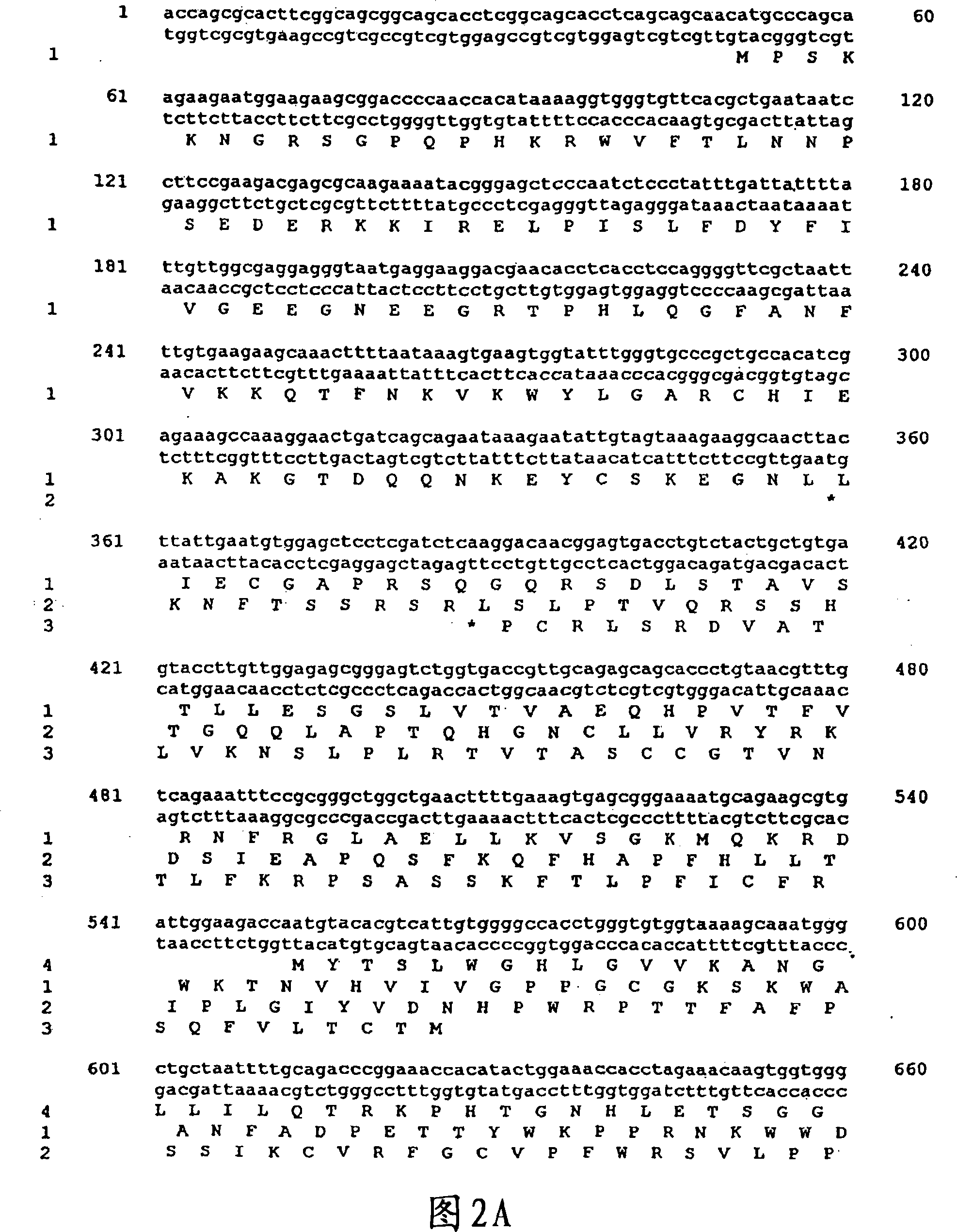
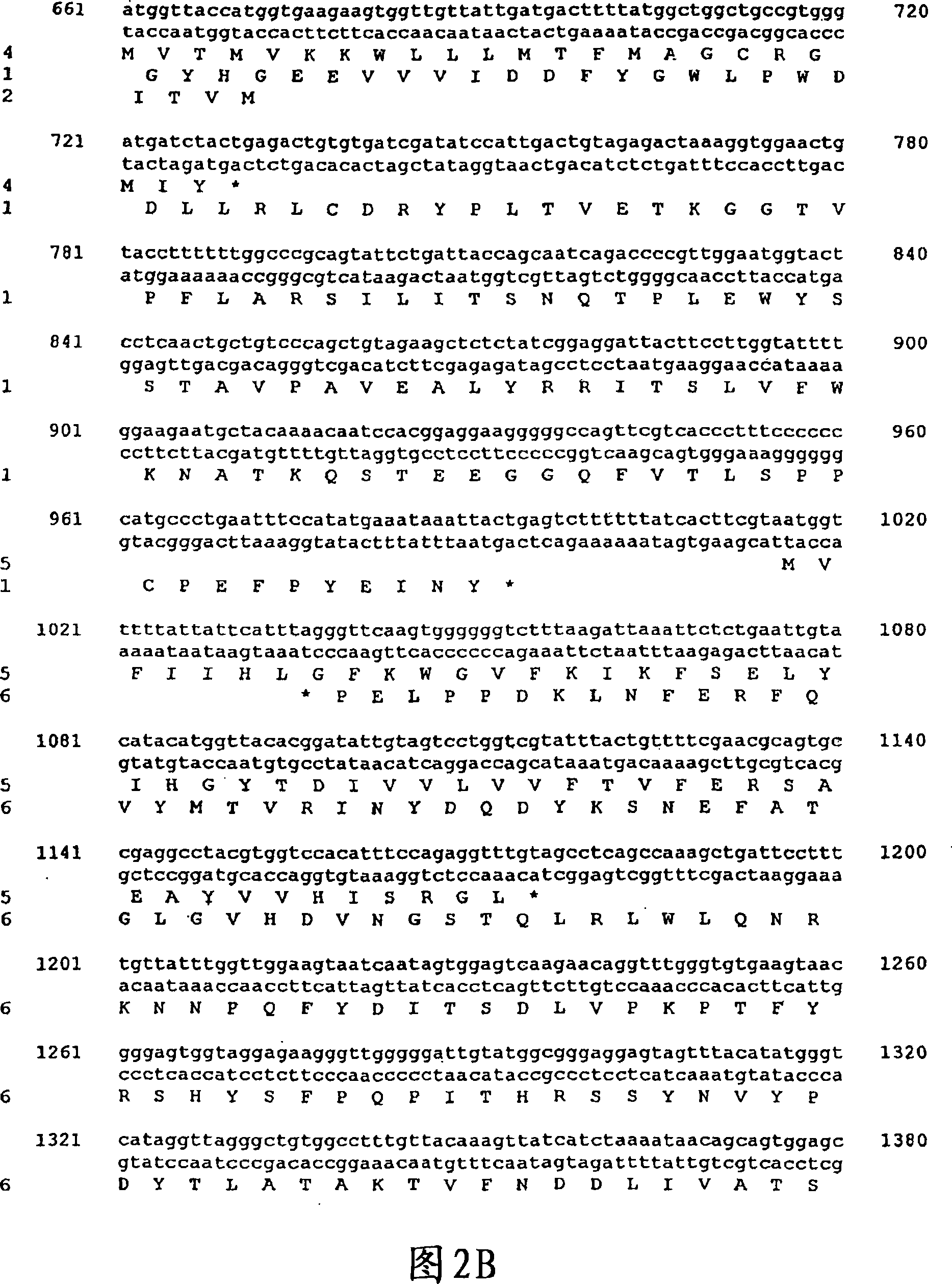
[0246] Porcine Circovirus: Conventional PCVI was isolated from persistently infected PK15 cells (ATCC CCL33). Isolate PCVII 412 was obtained from piglet lymph nodes challenged with lymph node homogenate from PMWS-affected piglets. This challenged piglet was diagnosed with PMWS. Isolate PCVII 9741 was isolated from the peripheral buffy coat of PMWS-affected piglets from the same herd following the isolation of PCVII 412. Isolate PCVII B9 was isolated from affected piglets in an American herd with a clinical outbreak of PMWS in the fall of 1997. ...
Embodiment 2
[0260] Example 2: PMWS reproduction
[0261] PMWS has not yet reproduced under controlled conditions, nor has its etiology been studied. To determine the causative agent of this disease, a number of tissues were collected from PMWS-affected pigs and studied as described in Example 1 above. Lymph nodes showed the most obvious gross lesions, histopathological changes, and circovirus infection was confirmed by immunostaining. Therefore, lymph nodes were used in the challenge experiments described above.
[0262] Challenge experiments performed as described in Materials and Methods successfully produced PMWS in pigs. In particular, some piglets died from infection and asymptomatic infected piglets developed PMWS-like microscopic lesions at the end of the experiment.
[0263] In another challenge experiment, the starting material used was lung tissue from pigs with chronic wasting and lymphadenopathy. These clinical signs are characteristic of PMWS. The tissue was combined w...
Embodiment 3
[0271] Example 3: Isolation and Propagation of PCVII
[0272] To determine the presence of infectious causative agents for PMWS, various tissues from pig #412 (experimentally challenged piglets sacrificed 21 days after infection) were used for virus isolation. Virus accumulation or adaptation was observed after serial passage of lymph node samples from pig #412 in Dulac cells. A distinct pattern of cytopathic effect initially developed, followed by an increase in virus titer determined by ELISA using standard Berlin anti-PCV antibodies as described above.
[0273] The presence of circovirus in Dulac cells infected with isolate PCVII 412 was subsequently detected by electron microscopy. After 6 passages, viral structural proteins were consistently detectable using Western blot assays.
PUM
 Abstract
Description
Claims
Application Information
 - R&D
- Intellectual Property
- Life Sciences
- Materials
- Tech Scout
- Unparalleled Data Quality
- Higher Quality Content
- 60% Fewer Hallucinations
Browse by: Latest US Patents, China's latest patents, Technical Efficacy Thesaurus, Application Domain, Technology Topic, Popular Technical Reports.
© 2025 PatSnap. All rights reserved.Legal|Privacy policy|Modern Slavery Act Transparency Statement|Sitemap|About US| Contact US: help@patsnap.com


