Whole blood preparation for cytometric analysis of cell signaling pathways
A cell counting and cell technology, applied in the analysis of materials, biomaterials analysis, individual particle analysis, etc.
- Summary
- Abstract
- Description
- Claims
- Application Information
AI Technical Summary
Problems solved by technology
Method used
Image
Examples
preparation example Construction
[0020] The present invention relates to a preparation method of a biological sample for the measurement of protein epitopes, which makes it possible to detect signal transduction pathways based on the ability to capture intracellular protein epitopes and transient activation states of epitopes. The methods provided by the present invention allow rapid fixation of biological samples containing erythrocytes to ensure that epitopes of signal transduction molecules and other intracellular protein epitopes remain in an activated state. The method of the present invention further allows lysis of red blood cells, thus making it a useful method for cytometric analysis of biological samples including, for example, whole blood, bone marrow aspirate, peritoneal fluid, and other red blood cell-containing sample. The present invention also provides a means to restore or "expose" epitopes on intracellular antigens that are cryptic due to the cross-linked fixative required to fix the sample....
Embodiment 1
[0062] Lysis and fixation of red blood cells and permeabilization of white blood cells
[0063] This example describes and compares the preparation of whole blood samples by hypotonic lysis followed by fixation and post-fixation detergent lysis.
[0064] Briefly, phorbol myristoyl ethyl ester (PMA, Sigma Chemical Corp., St. Louis, MO) was prepared as a 40 M working solution in 100% absolute ethanol and used in whole blood at a final concentration of 400 nM . Triton X-100 and other detergents used in this example were prepared in Surfact-Pak TM Detergent sample kit formats were purchased from Pierce Biotechnology (Rockford, IL). The sample kit contains seven different nonionic detergents (Tween-20, Tween-80, Triton X-100, Triton X-114, Nonidet P-40, Brij-35 and Brij-58, all named 10% aqueous solution) and three powders (nonionic octyl-B-glucoside, and octyl-B-thioglucopyranoside, and amphoteric CHAPS). The powdered detergent was dissolved in PBS (without Ca 2+ and Mg 2+ ...
Embodiment II
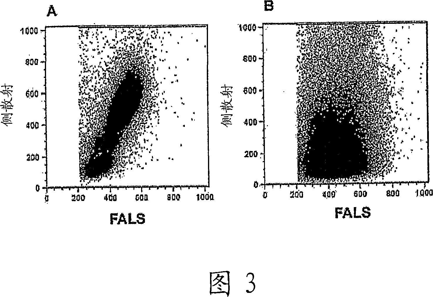
[0089] Effects of Fixative and Detergent Concentrations on Leukocyte Light Scattering
[0090] This example demonstrates the effect of concentration and incubation time of both fixative and detergent on the degree of separation and recovery of leukocytes.
[0091] The effect of different fixative concentrations was investigated following previous studies suggesting that high concentrations of cross-linked fixatives help maintain the light-scattering profile and separation of leukocyte populations. Whole blood samples were incubated in increasing concentrations of formaldehyde (from 1% to 10% final concentration) for 10 minutes at room temperature or 37°C, followed immediately by incubation with 1 ml of 0.1% TX-100 (at room temperature). As shown in Figure 6, gradually increasing the concentration of formaldehyde from 2% to 4% (for room temperature incubation) significantly improved the separation of leukocyte populations (left panel). Similarly, treatment of whole blood sam...
PUM
| Property | Measurement | Unit |
|---|---|---|
| time | aaaaa | aaaaa |
| time | aaaaa | aaaaa |
| volume | aaaaa | aaaaa |
Abstract
Description
Claims
Application Information
 - R&D
- Intellectual Property
- Life Sciences
- Materials
- Tech Scout
- Unparalleled Data Quality
- Higher Quality Content
- 60% Fewer Hallucinations
Browse by: Latest US Patents, China's latest patents, Technical Efficacy Thesaurus, Application Domain, Technology Topic, Popular Technical Reports.
© 2025 PatSnap. All rights reserved.Legal|Privacy policy|Modern Slavery Act Transparency Statement|Sitemap|About US| Contact US: help@patsnap.com


