
Fluoroscopic image early cancer diagnosis equipment

A fluorescent image and diagnostic instrument technology, applied in the field of medical instruments, can solve the problems that it is difficult to ensure that the target illumination is in good condition, affects the accurate diagnosis of malignant tumors, and is difficult to operate by hand.

Active Publication Date: 2009-08-19
衍莹生物科技(苏州)有限公司
View PDF3 Cites 0 Cited by
  • Summary
  • Abstract
  • Description
  • Claims
  • Application Information

AI Technical Summary

Problems solved by technology

However, in this patent, the tumor tissue illumination and image acquisition system is difficult to design very small, and there are no forceps holes and bending control devices for auxiliary channels like the current medical endoscope, so when making a diagnosis of narrow organs in the body It will bring a lot of inconvenience and difficulties; in this patent technology, three independent imaging CCDs are used, as well as a whole set of optical imaging elements and adjustment systems. The way is to intercept still images for display to make a clear diagnosis. This method of intercepting still images is difficult to ensure that the target illumination is in good condition when the relative distance and position between the probe and the target change randomly, making it difficult to control the quality of image capture, which will seriously Accurate diagnosis of malignant tumors

Method used


Image

  • Fluoroscopic image early cancer diagnosis equipment
  • Fluoroscopic image early cancer diagnosis equipment
  • Fluoroscopic image early cancer diagnosis equipment

Examples


Embodiment Construction

[0027] The specific implementation manner of the present invention will be further introduced below.

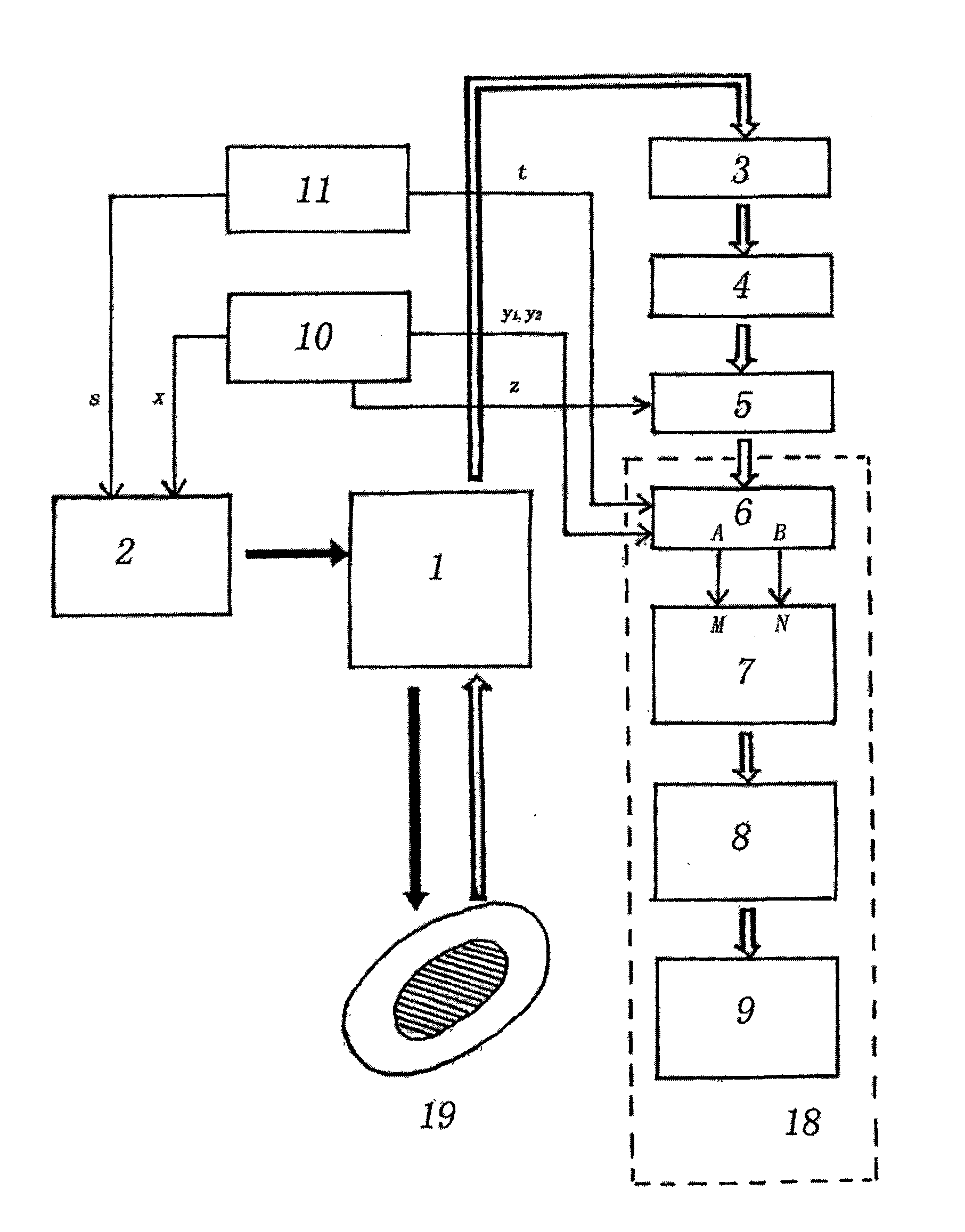
[0028] The traditional Chinese medical endoscope (1) of the present invention adopts domestic model and is XS-30 fiber gastroscope.

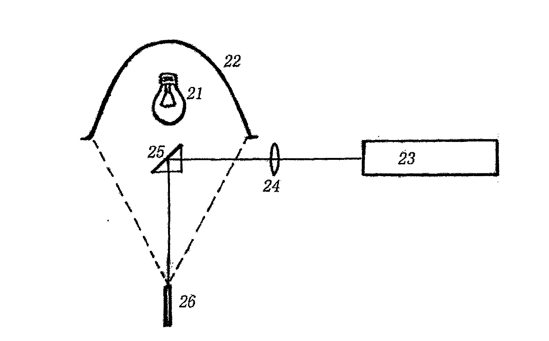
[0029] The mixed light source in the light source adapter (2) adopts a halogen lamp (150W) and a 405nm semiconductor laser light source (above 50mw), such as figure 2 shown. The halogen lamp (21) is placed on the first focal point of the ellipsoidal condenser (22) with a light-condensing function, and the light inlet port (26) of the medical endoscope light guide is placed on the second focal point of the ellipsoidal condenser (22), in order to To make the laser beams mix without hindrance, the distance between the two focal points of the ellipsoid mirror is not less than 70mm. After being focused, the light beam output by the laser (23) is refracted at 90° by the 8×8mm total reflection shuttle mirror and enters the light guide port (26) of ...


PUM


Abstract

The present invention is a fluorescence image early cancer diagnostic instrument. The present invention adopts a dynamic and real-time display method of fluorescence images, which not only has extremely high spatial resolution, but also has extremely high time resolution, and can collect all the fluorescence image information of the diagnosis area at any time. , without missing any detail. It is much superior to the method of capturing still images for diagnosis, and it also greatly avoids errors and missed detections caused by not capturing still images in time and accurately, thereby greatly improving the accuracy of early cancer detection. The invention has the advantages of accuracy, intuition, no side effect, simple operation method and the like, and can be used for diagnosing various malignant tumors in vivo and in vitro. Moreover, it can help determine the scope of the operation before the operation, and can also be followed up during the operation to judge the effect of the operation and provide instructions for timely removal of residual tumors.

Description

Technical field: [0001] The invention belongs to the technical field of medical instruments, and relates to a fluorescent image early cancer diagnostic instrument. Background technique: [0002] Now the fluorescence diagnostic instrument for malignant tumors uses the tumor tissue to generate fluorescence under the excitation of a certain wavelength of light, and then observes the fluorescence image after receiving the fluorescence of the tumor tissue, and distinguishes the diagnosis from the different colors in the fluorescence image. For example, on December 5, 2002, Fudan University and Renji Hospital Affiliated to Shanghai Second Medical University jointly applied for the patent of "Malignant Tumor Fluorescent Image Positioning Diagnostic Apparatus". [0003] The fluorescent image positioning diagnostic instrument for malignant tumors uses human tissue to generate fluorescence under the excitation of light of a certain wavelength. The diagnostic instrument formed by the ...

Claims


Application Information

Patent Timeline
Patent Type & Authority Patents(China)
IPC IPC(8): A61B5/00A61B1/04
Inventor 叶衍铭萧树东
Owner 衍莹生物科技(苏州)有限公司
Learn More