hmg-1 gene nucleic acid in situ hybridization detection kit, detection method and application
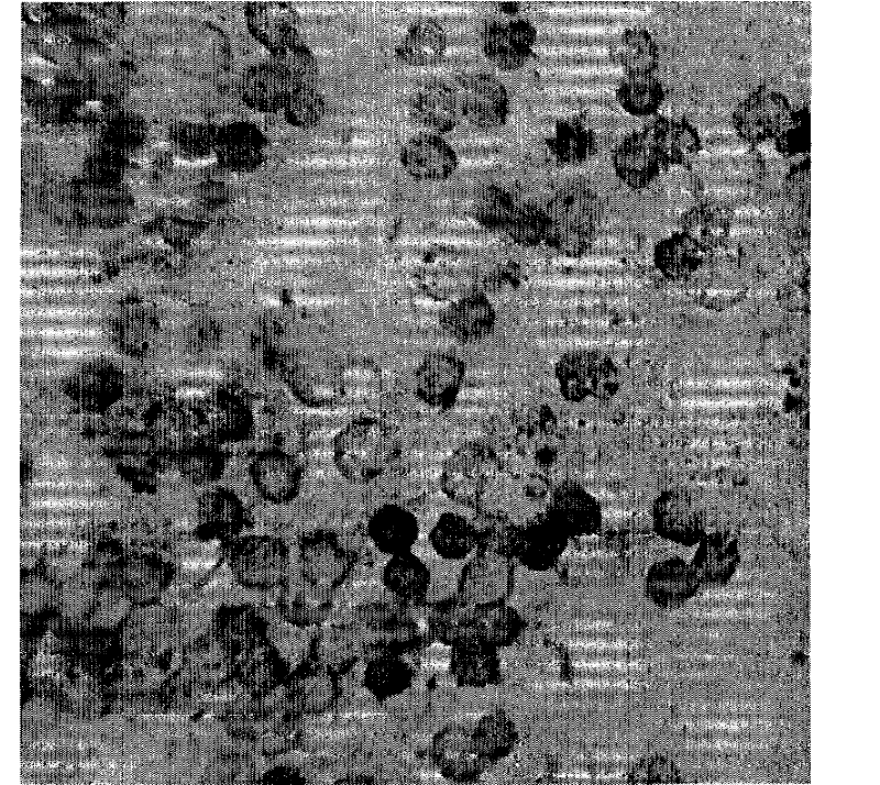
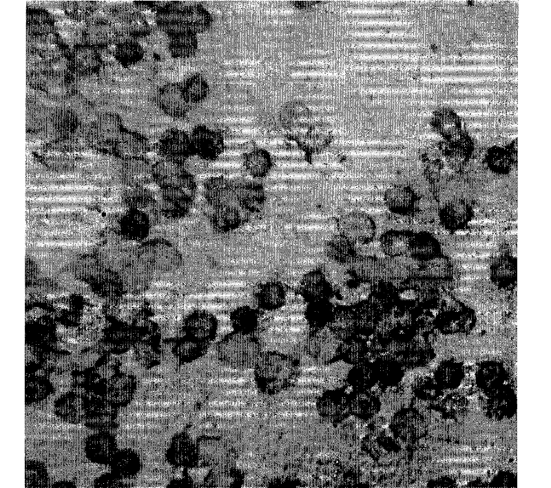
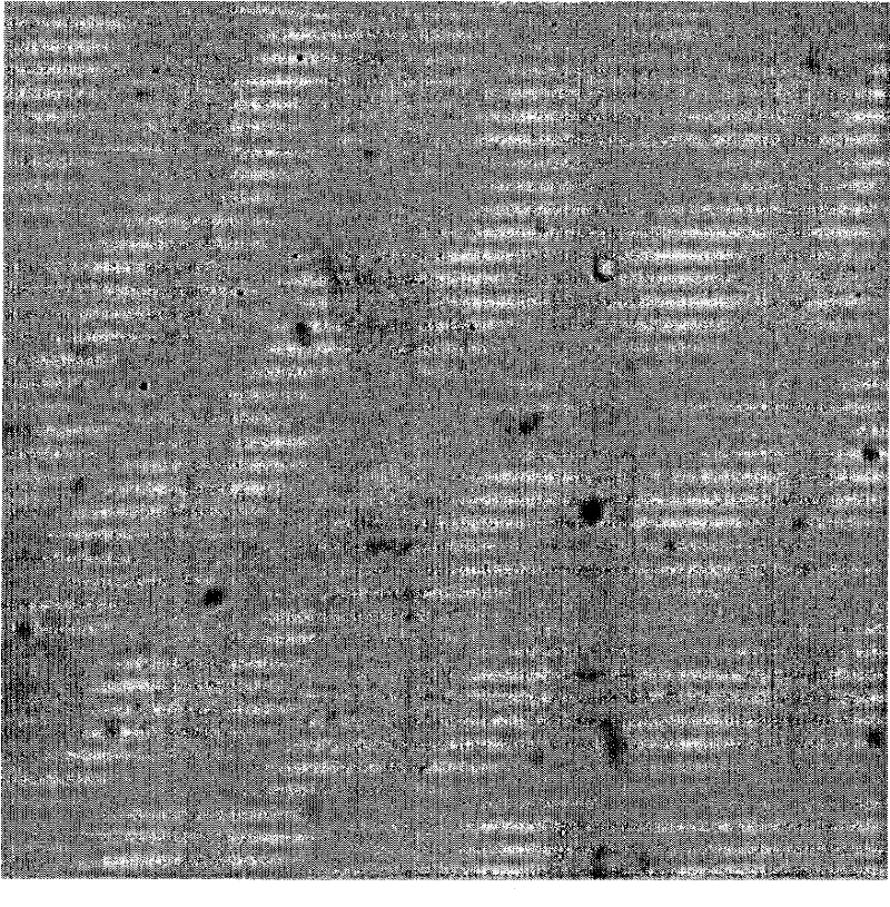
A detection kit and in situ hybridization technology, applied in the field of cancer gene detection technology, can solve problems such as drug resistance of tumor cells, unreported HMG-1 gene detection technology, failure of the anti-cancer war, etc., to reduce mortality and the effect on morbidity
- Summary
- Abstract
- Description
- Claims
- Application Information
AI Technical Summary
Problems solved by technology
Method used
Image
Examples
Embodiment 1
[0039] Prepare the in situ hybridization kit of this embodiment according to conventional methods, the kit includes hybridization probes and markers designed with the HMG-1 gene as the detection target gene, wherein:
[0040] Digoxigenin was selected as the probe label in this embodiment.
[0041] Kit composition:
[0042] Digestive solution 100μL / tube 1 tube / box Colorless transparent liquid
[0043] Protective solution 100μL / tube 1 tube / box Colorless transparent liquid
[0044] Pre-hybridization solution 1300μL / tube 2 tubes / box Colorless transparent liquid
[0045] Sense hybridization solution 10μL / tube 1 tube / box Colorless transparent liquid
[0046] Antisense hybridization solution 10μL / tube 1 tube / box Colorless transparent liquid
[0047] Blocking solution 1000μL / tube 1 tube / box Colorless transparent liquid
[0048] Alkaline phosphatase antibody 1μL / tube 1 tube / box Colorless transparent liquid
[0049] Chromogen A 175μL / tube 1 tube / box Yellow liquid
[0050] Chromog...
Embodiment 2
[0091] Specimen processing:
[0092] 1). Use a 10ml centrifuge tube to fill 4.5ml of lymphocyte separation solution, then slowly add 3ml of anticoagulated blood into the centrifuge tube containing lymphocyte separation solution (blood: lymphocyte separation solution = 1:1.5), 2000r / min Centrifuge for 10min
[0093] 2). Draw the white blood cells in the middle layer into another centrifuge tube, then add about twice the amount of 1× buffer I to this tube, mix well, and centrifuge at 1500g / min for 10min
[0094] 3). Discard the supernatant. Add about twice the 1× buffer I to the pellet, mix well, and centrifuge at 1500g / min for 10min
[0095] 4). Discard the supernatant, and absorb the excess liquid from the mouth of the test tube with paper towels. Then the precipitate was made into a suspension, dropped on a glass slide, and allowed to dry naturally. (Hospitals with conditions can use the film-making machine to make films.) With 3ml of blood, 4 films can be made.
[0096] ...
Embodiment 3
[0099] Prepare the reagents in the kit to the concentration used
[0100] 1). Dilute 10× buffer I with triple distilled water at 1:10 to 1× buffer I;
[0101] 2). Dilute 20× buffer II with triple distilled water at 1:10 to 2× buffer II;
[0102] Dilute 1:100 into 0.2×buffer II; dilute 1:200 into 0.1×buffer II;
[0103] 3). Dilute 10× buffer III with triple distilled water at 1:10 to 1× buffer III;
[0104] 4). Dilute 10× buffer IV with triple distilled water at a ratio of 1:10 to form × buffer IV (take 10 mL each of 1#, 2#, and 3#, and add water to 100 mL).
PUM
 Abstract
Description
Claims
Application Information
 - Generate Ideas
- Intellectual Property
- Life Sciences
- Materials
- Tech Scout
- Unparalleled Data Quality
- Higher Quality Content
- 60% Fewer Hallucinations
Browse by: Latest US Patents, China's latest patents, Technical Efficacy Thesaurus, Application Domain, Technology Topic, Popular Technical Reports.
© 2025 PatSnap. All rights reserved.Legal|Privacy policy|Modern Slavery Act Transparency Statement|Sitemap|About US| Contact US: help@patsnap.com


