
Non-invasive fetal sex determination

a fetal sex and non-invasive technology, applied in the field of non-invasive sex determination, can solve the problem of inherently inefficient random or shotgun sequencing methods

Active Publication Date: 2018-06-12
ROCHE MOLECULAR SYST INC
View PDF227 Cites 0 Cited by
  • Summary
  • Abstract
  • Description
  • Claims
  • Application Information

AI Technical Summary

Benefits of technology

[0057]The assay methods of the invention include selective enrichment of selected nucleic acid regions from the Y chromosome and one or more non-Y reference chromosomes. A distinct advantage of the invention is that the selected nucleic acid regions can be further analyzed using a variety of detection and quantification techniques, including but not limited to hybridization techniques, digital PCR, and, preferably, high-throughput sequencing determination techniques. Primers can be designed against any number of selected nucleic acid regions for any chromosome in addition to the Y chromosome. Although amplification prior to the identification and quantification of the selected nucleic acid regions is not mandatory, limited amplification prior to detection is preferred.
[0058]The present invention is an improvement over more random techniques such as massively parallel shotgun sequencing (e.g., random sequencing) or the use of random digital PCR that have been used recently to detect copy number variations in maternal samples such as maternal blood. The aforementioned approach relies upon sequencing of all or a statistically significant population of DNA fragments in a sample, followed by mapping of or otherwise associating the fragments to their appropriate chromosomes. The identified fragments are then compared against each other or against some other reference (e.g., a sample with a known normal chromosomal complement) to determine copy number variation of particular chromosomes. Random or shotgun sequencing methods are inherently inefficient as compared to the present invention, as the data generated on the chromosomal regions of interest constitute only a minority of the data that is generated.
[0059]Techniques that are dependent upon a very broad sampling of DNA in a sample provide a broad coverage of the DNA analyzed, but in fact are sampling the DNA contained within a sample on a 1× or less basis (i.e., subsampling). In contrast, the selective amplification and / or enrichment techniques (such as hybridization) used in the present methods provide depth of coverage of only the selected nucleic acid regions; and as such provide a “super-sampling” of the selected nucleic acid regions with an average sequence coverage of preferably 2× or more, more preferably sequence coverage of 100× of more, 200× or more, 250× or more, 500× or more, 750× or more or even more preferably sequence coverage of 1000× or more of the selected nucleic acid regions.
[0060]Thus, the substantial majority of sequences analyzed for quantification of Y chromosomal sequences are informative of the presence of one or more selected nucleic acid regions on the Y chromosome and one or more non-Y chromosomes. The methods of the invention do not require analysis of large numbers of sequences that are not from the chromosomes of interest and that do not provide information on the relative quantity of the chromosomes of interest.Detecting and Quantifying the Y Chromosome
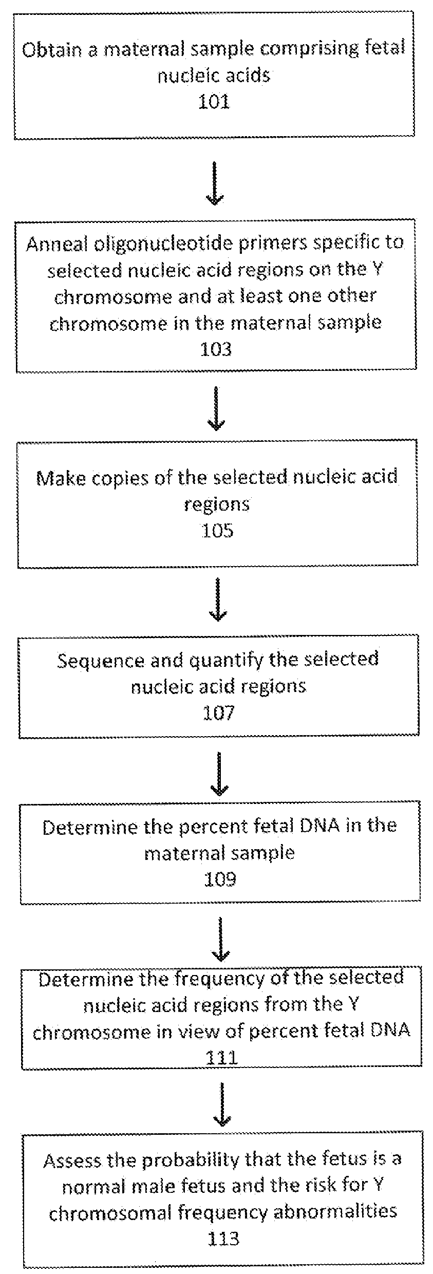
[0061]The present invention provides methods for determining fetal sex, and / or for identifying Y chromosomal aneuploidies or sex chromosome mosaicisms, and / or for determining possible sample contamination in maternal samples. The samples are maternal samples comprising both maternal and fetal DNA such as maternal blood samples (i.e, whole blood, serum or plasma). The methods enrich and / or isolate and amplify one or, preferably, several to many selected nucleic acid regions in a maternal sample that correspond to the Y chromosome and one or more reference (non-Y) chromosomes which are used to determine the presence or absence and relative quantity or frequency of Y chromosomal sequences in view of the percent of fetal DNA present in the sample. As described in detail supra, the methods of the invention preferably employ one or more selective amplification cycles (e.g., using one or more primers that specifically hybridize to the selected nucleic acid regions) or enrichment (e.g., hybridization and separation) steps to enhance the content of the selected nucleic acid regions in the sample. The selective amplification and / or enrichment steps typically include mechanisms to engineer copies of the selected nucleic acid regions for further isolation, amplification and analysis. This selective approach is in direct contrast to the random amplification approach used by other techniques, e.g., massively parallel shotgun sequencing, as such techniques generally involve random amplification of all or a substantial portion of the genome.
[0062]One challenge with the detection and quantification of Y chromosome-specific sequences in a maternal sample is that the majority of the cell free fetal DNA as a percentage of total cell free DNA in a maternal sample such as blood serum or plasma may vary from less than one to about forty percent, and most commonly the percentage of fetal DNA in a maternal sample is below twenty percent and frequently at or below ten percent. Thus, for example, in a maternal sample that is 10% fetal DNA, each chromosome will contribute 1 / 46th of 10% (or approximately 0.22%) in a normal fetus. In a male fetus, the Y chromosome will thus contribute 1 / 46th of the 10% (0.22%) and autosomes will contribute 2 / 46th or 1 / 23rd of 10% (0.44% as there are two of each autosome). Thus, in determining whether a fetus is a normal male fetus, the frequency of Y chromosome-specific sequences in a sample that is 10% fetal should be 0.22% and the frequency of, for example, chromosome 3-specific sequences should be 0.44% since a male fetus has two chromosome 3s. In determining whether there is a Y chromosomal aneuploidy (that is, two or more Y chromosomes), the frequency of Y chromosome-specific sequences would be approximately 0.44% or more (i.e., 0.66% for a trisomy Y). In determining whether a fetus may be a sex chromosome mosaic, the frequency of Y chromosome-specific sequences should be less and may be substantially less than 0.22%, and the same would be true for assessing the likelihood of sample contamination of a maternal sample with nucleic acids from a female fetus contaminated by a maternal sample with nucleic acids from a male fetus. If one is to detect specific nucleic acids present at such a low percentage robustly through the methods described herein, the variation in the measurement of the extra chromosome has to be significantly less than the percent increase of the extra chromosome.

Problems solved by technology

Random or shotgun sequencing methods are inherently inefficient as compared to the present invention, as the data generated on the chromosomal regions of interest constitute only a minority of the data that is generated.

Method used


Image

  • Non-invasive fetal sex determination
  • Non-invasive fetal sex determination
  • Non-invasive fetal sex determination

Examples


example 1

Detection of Y Chromosome Frequency Abnormalities Using Non-polymorphic Sites in Chromosome-specific Genomic Regions

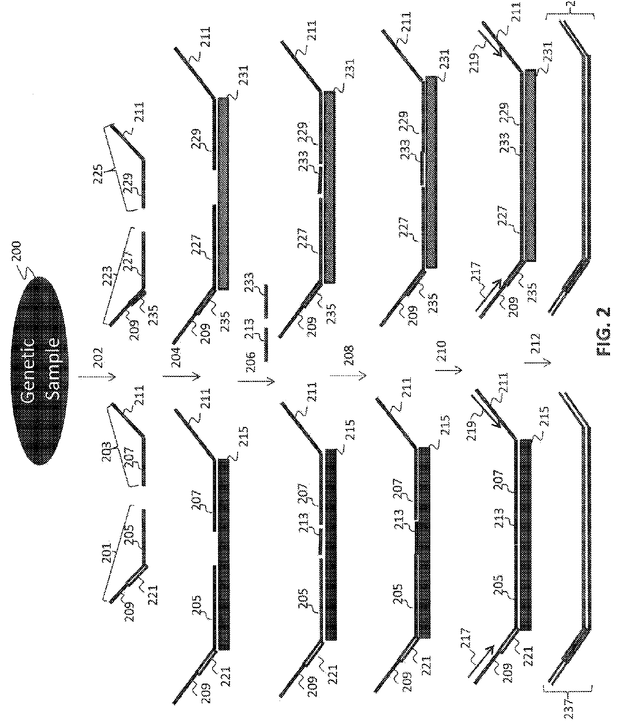
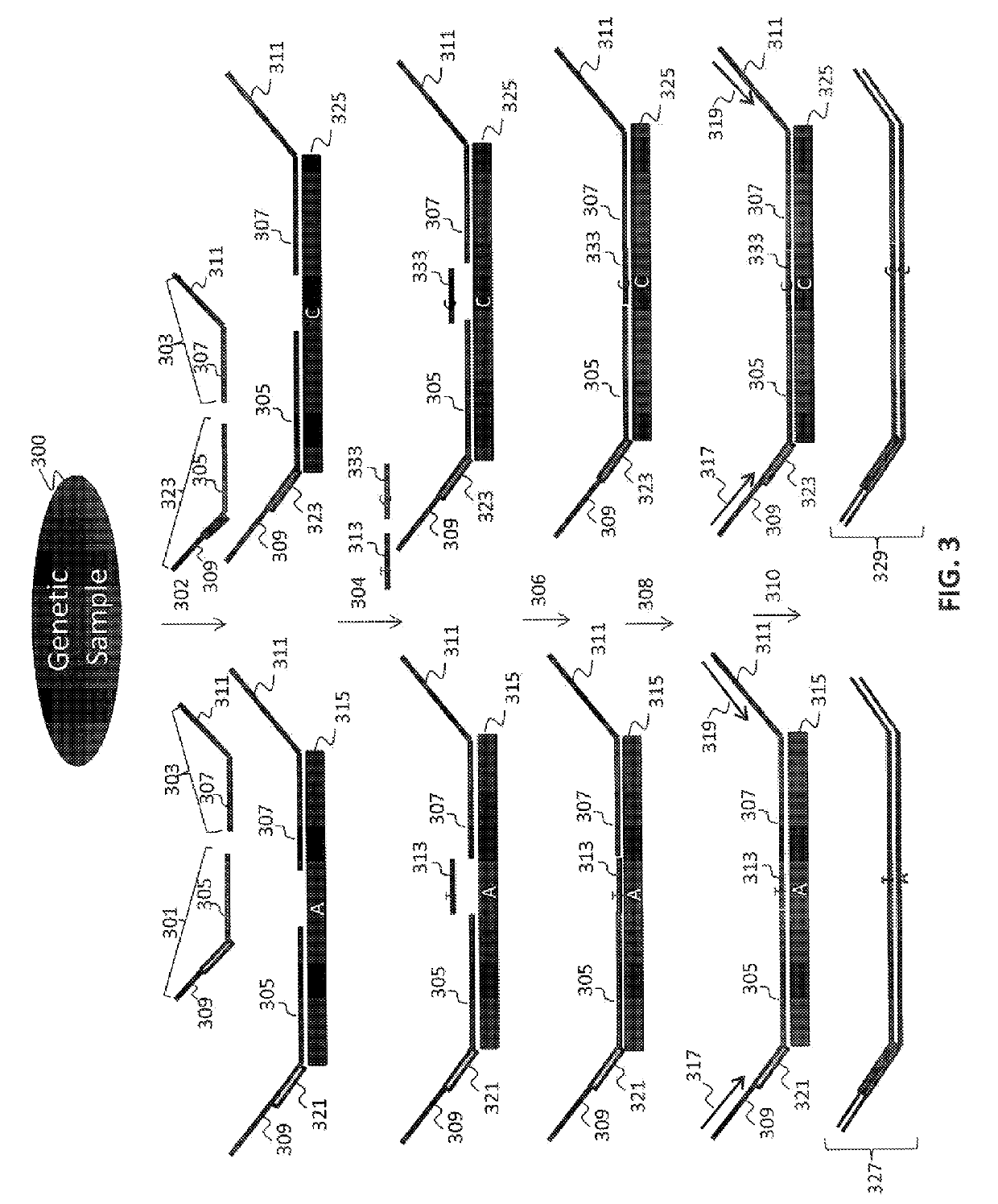
[0156]In a first embodiment, assays directed against a specific genomic regions on the Y chromosome were used to identify the presence or absence of a Y chromosome frequency abnormality. The present assay system allowed the identification of the presence or absence of such an abnormality in the DNA of multiple individuals using a highly multiplexed system.

[0157]Multiple interrogations were prepared using oligonucleotides complementary to or derived from the Y chromosome (chrY), and chromosomes 13, 18 and 21 (chr13, chr18 and chr21). All oligonucleotides used in the tandem ligation formats were synthesized using conventional solid-phase chemistry. The oligos of the first fixed set and the bridging oligonucleotides were synthesized with 5′ phosphate moieties to enable ligation to 3′ hydroxyl termini of adjacent oligonucleotides. Thirty-two non-polymorphic assays were dev...

example 2

Preparation of DNA for Use in Tandem Ligation Procedures

[0158]Genomic DNA from subjects was obtained from Coriell Cell Repositories (Camden, ew Jersey) and fragmented by acoustic shearing (Covaris, Woburn, Mass.) to a mean fragment size of approximately 200 bp.

[0159]The DNA was biotinylated using standard procedures. Briefly, the Covaris fragmented DNA was end-repaired by generating the following reaction in a 1.5 ml microtube: 5 ug DNA, 12 μl 10× T4 ligase buffer (Enzymatics, Beverly Mass.), 50 U T4 polynucleotide kinase (Enzymatics, Beverly Mass.), and H20 to 120 μl. This was incubated at 37° C. for 30 minutes. The DNA was diluted using 10 mM Tris 1 mM EDTA pH 8.5 to desired final concentration of ˜0.5 ng / μl.

[0160]5 μl DNA was placed in each well of a 96-well plate, and the plate sealed with an adhesive plate sealer and spun for 10 seconds at 250×g. The plate was then incubated at 95° C. for 3 minutes, and cooled to 25° C., and spun again for 10 seconds at 250×g. A biotinylation m...

example 3

Universal Amplification of Ligated Products

[0166]The polymerized and / or ligated nucleic acids were amplified using universal PCR primers complementary to the universal sequences present in the first and second fixed sequence oligos hybridized to the nucleic acid regions of interest. 25 μl of each of the reaction mixtures of Example 3 were used in each amplification reaction. A 50 μL universal PCR reaction consisting of 25 μL eluted ligation product plus 1×Pfusion buffer (Finnzymes, Finland), 1M Betaine, 400 nM each dNTP, 1 U Pfusion error-correcting thermostable DNA polymerase, and primer pairs with sample tags used to uniquely identify individual samples prior to pooling and sequencing. The PCR was carried out under stringent conditions using a BioRad Tetrad™ thermocycler.

[0167]10 μl of universal PCR product from each of the samples were pooled and the pooled PCR product was purified using AMPure™ SPRI beads (Beckman-Coulter, Danvers, Mass.), and quantified using Quant-iT™ PicoGree...


PUM

PropertyMeasurementUnit
concentrationaaaaaaaaaa
v/vaaaaaaaaaa
pHaaaaaaaaaa

Abstract

The present invention provides methods for non-invasive determination of sex in a fetus or of Y chromosomal frequency abnormalities—indicative of aneuploidy or sex mosaicisms in a fetus—by detecting and determining the relative contribution genetic sequences from the Y chromosome in view of the percent fetal contribution in a maternal mixed sample.

Description

FIELD OF THE INVENTION[0001]This invention relates to methods for non-invasive sex determination of a fetus or of Y chromosomal frequency abnormalities by detecting and determining the relative contribution of genetic sequences from the Y chromosome in view of the percent fetal contribution in a maternal mixed sample.BACKGROUND OF THE INVENTION[0002]In the following discussion certain articles and methods will be described for background and introductory purposes. Nothing contained herein is to be construed as an “admission” of prior art. Applicant expressly reserves the right to demonstrate, where appropriate, that the articles and methods referenced herein do not constitute prior art under the applicable statutory provisions.[0003]Genetic abnormalities account for a wide number of pathologies, including syndromes caused by chromosomal aneuploidy (e.g., Down syndrome) and those caused by germline mutations resulting in either monogenic or polygenic diseases or disorders. Detection ...

Claims


Application Information

Patent Timeline
Patent Type & Authority Patents(United States)
IPC IPC(8): C12Q1/68
CPCC12Q1/6883C12Q1/6879C12Q2600/156
Inventor STRUBLE, CRAIGOLIPHANT, ARNOLDWANG, ERIC
Owner ROCHE MOLECULAR SYST INC
Learn More