Specific biomarker set for non-invasive diagnosis of liver cancer

a biomarker and liver cancer technology, applied in the field of specific biomarker sets for non-invasive diagnosis of liver cancer, can solve the problems of high mortality rate, afp test, excessive false positive, etc., and achieve the effect of higher fluorescent intensity and higher expression of the corresponding biomarkers

Active Publication Date: 2018-02-06
DRAGON VICTORY DEV
View PDF30 Cites 1 Cited by
  • Summary
  • Abstract
  • Description
  • Claims
  • Application Information

AI Technical Summary

Benefits of technology

[0015]In the present invention, a detection and quantification method measuring the auto-antibodies against a list of specific tumor biomarker aiming for diagnosing and staging cancers is provided. Comparing to the normal liver epithelial cells, HCC tumor cells tend to produce a unique set of proteins. The evaluation of the unique protein set, biomarkers, will complement the conventional diagnostic methods and facilitate early detection of cancers.
[0017]Specificity and accuracy of this set of liver cancer biomarkers are then validated and taken together for diagnosis of liver cancers. In the present invention, proteins of the listed biomarkers are expressed from cDNA clones, purified and coupled to fluorescent microsphere beads with different emission wavelengths. Auto-antibodies present in patients' sera against the proteins immunologically bind to the protein-bead conjugate. The auto-antibodies subsequently interact with PE-conjugated secondary antibodies. The specific fluorescence signal of the microsphere beads serves as an identifier for the conjugated biomarkers. By measuring the fluorescent intensity given by the PE-conjugated secondary antibodies at the complex, it allows the detection and quantification of the auto-antibodies. Since the auto-antibodies are produced in the patients' sera in proportion to the abundance of the biomarkers at HCC tumor cells, the higher fluorescent intensity resulted from higher concentration of auto-antibodies indicates the higher expression of the corresponding biomarkers. The lowest detection limit of each biomarker to the total serum auto-antibodies is about 0.15 ng / mL.
[0018]Comparing to sera from healthy subjects, the level of auto-antibodies against the target biomarkers is at a higher concentration in cancer patient. Moreover, comparing different sera from liver cancer patients at different stages, signature patterns for staging may be generated. Thus, the present invention allows the non-invasive evaluation of the targeted liver cancer biomarker. This enables the detection of HCC at early stages and the identification of signature biomarker patterns for staging, as well as the detection of recurrences during a monitoring period of post-chemotherapeutic treatment.

Problems solved by technology

Most HCC patients have rapid tumor progressing resulting in high mortality rate.
However, the major problem of the AFP test is excessive false positive.
Despite AFP test is commonly suggested for diagnosis of liver cancers, its result is not conclusive.
However, conventional detection of HCCs comes with several limitations: (a) About 20% of liver cancers does not produce elevated level of the commonly used HCC tumor markers [2].
(c) Ultrasound is not able to detect small tumors [4].
(e) MRI scans are expensive and the procedure is time consuming.
However, not all biomarkers themselves can be found in serum or urine for convenient diagnosis.
Ideally, examination of auto-antibodies from peripheral blood would be a testament for detecting liver cancers early, and in a non-invasive manner One common hurdle hindering clinical use of biomarkers is that they have not been validated after discovery.
But once validated, such test would be cost effective and accurate.

Method used


Image

  • Specific biomarker set for non-invasive diagnosis of liver cancer
  • Specific biomarker set for non-invasive diagnosis of liver cancer
  • Specific biomarker set for non-invasive diagnosis of liver cancer

Examples


example 1a

Protein Extraction from Patients' Biopsies

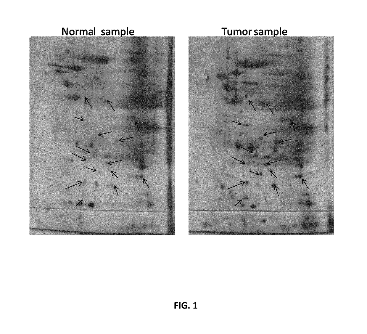
[0060]500 mg of the paired patients' biopsies (tumor biopsy versus juxtaposed normal tissue) are collected and washed with PBS. The tissues are frozen by submerging into liquid nitrogen and immediately homogenized with pestle and mortar. To the homogenized samples, lysis solution (8M Urea, 4% CHAPS, 2% IPG Buffer, 0.2 mg / ml PMSF) is added, then vortex for at least 5 min until the tissues are completely dispersed. The lysates are then clarified by centrifugation at 14,000 rpm for 10 minutes at 4° C. The supernatants are further cleaned up by 2D Clean Up kit (Amersham) to remove the salt and impurities. The pellets are resuspended with minimum volume of Rehydration Solution (No DTT & IPG Buffer added). The protein concentrations are then measured by Bio-Rad protein assay and aliquots of 200 g / per tube are stored at −70° C.

example 1b

Resolving Proteins by Two-Dimensional Electrophoresis

[0061]To 1 ml rehydration stock solution, 2.8 mg DTT, 5 μl pharmalyte or IPG Buffer, and 2 μl bromophenol blue are added. 50-100 μg of protein sample is added to the 13 cm Immobiline DryStrip (IPG strip) containing 250 μl of rehydration solution. After removing the protective cover, the IPG strip is positioned in the strip holder with the gel side facing down, and overlaid with Cover Fluid to prevent dehydration during electrophoresis. The strip is then placed on to Ettan IPGphor (Amersham) for isoelectric focusing (first dimensional electrophoresis).

[0062]After the first-dimensional electrophoresis, the IPG strip is equilibrated with equilibrate solution (6 M Urea 2% SDS, 50 mM Tris HCl pH 6.8, 30% Glycerol, 0.002% Bromophenol blue, 100 mg DTT per 10 ml buffer and 250 mg IAA per 10 ml buffer), and then washed with 1×SDS running Buffer for 4-5 times. The IPG strip is placed on top of the second-dimension gel and overlaid with seal...

example 2a (

SEQ ID NO.1)

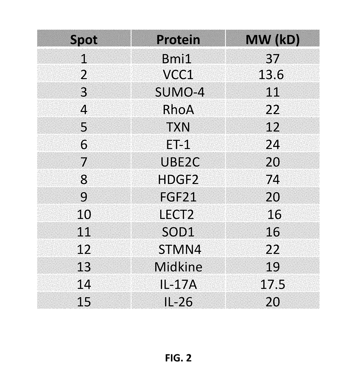
Amino Acid Sequence of Bmi1

[0064]

MHRTTRIKITELNPHLMCVLCGGYFIDATTIIECLHSFCKTCIVRYLETSKYCPICDVQVHKTRPLLNIRSDKTLQDIVYKLVPGLEKNEMKRRRDFYAAHPSADAANGSNEDRGEVADEDKRIITDDEIISLSIEFFDQNRLDRKVNKDKEKSKEEVNDKRYLRCPAAMTVMHLRKFLRSKMDIPNTFQIDVMYEEEPLKDYYTLMDIAYIYTWRRNGPLPLKYRVRPTCKRMKISHQRDGLTNAGELESDSGSDKANSPAGGIPSTSSCLPSPSTPVQSPHPQFPHISSTMNGTSNSPSGNHQSSFANRPRKSSVNGSSATSSG


PUM

PropertyMeasurementUnit
pHaaaaaaaaaa
total volumeaaaaaaaaaa
temperatureaaaaaaaaaa

Abstract

Cells within liver tumour mass comprise a unique set of proteins / tumour antigens when compared to the normal liver tissues epithelial cells juxtaposed to the tumour. The presence of tumour antigens couples the production of auto-antibodies against these tumour antigens. The present invention relates to the identification and elucidation of a protein set that can act as a novel marker set for liver cancer diagnosis and prognosis. Specifically, it relates to a kit that enables diagnostic and prognostic measurement of auto-antibodies in serum of liver cancer patients. The present invention provides a non-invasive, specific, sensitive, and cost effective detection and quantification method by evaluating a set of validated liver cancer proteins / tumour antigens, which includes Bmi-1, VCC1, SUMO-4, RhoA, TXN, ET-1, UBE2C, HDGF2, FGF21, LECT2, SOD1, STMN4, Midkine, IL-17A or IL26, to complement the conventional diagnostic methods.

Description

COPYRIGHT NOTICE / PERMISSION[0001]A portion of the disclosure of this patent document contains material which is subject to copyright protection. The copyright owner has no objection to the facsimile reproduction by anyone of the patent document or the patent disclosure as it appears in the Patent and Trademark Office patent file or records, but otherwise reserves all copyright rights whatsoever. The following notice applies to the processes, experiments, and data as described below and in the drawings attached hereto: Copyright© 2014, Vision Global Holdings Limited, All Rights Reserved.TECHNICAL FIELD[0002]The present invention describes a detection and quantification method for a list of specific and novel Hepatocellular Carcinoma (HCC) tumor biomarkers, by measuring the corresponding auto-antibodies in liver cancer patients' sera. The set of biomarkers comprises Bmi1, VCC1, SUMO-4, RhoA, TXN, ET-1, UBE2C, HDGF2, FGF21, LECT2, SOD1, STMN4, Midkine, IL-17A and IL26. More specificall...

Claims


Application Information

Patent Timeline
Patent Type & Authority Patents(United States)
IPC IPC(8): G01N33/564G01N33/58G01N33/574
CPCG01N33/57438G01N33/582G01N33/564G01N2458/00
Inventor MAN, CORNELIA WING YINWAI, NORMAN FUNG MANWONG, BING LOUWAI, BENJAMIN CHI YIN
Owner DRAGON VICTORY DEV
Why Eureka
Try Eureka