Methods of inducing photosensitivity by targeting channelrhodopsin-2 and halorhodopsin to subcellular regions of retinal ganglion cells
a technology of halorhodopsin and channelrhodopsin, which is applied in the direction of antibody medical ingredients, peptide/protein ingredients, metabolism disorders, etc., can solve the problem of loss of visual information processing ability and achieve the effect of restoring sigh
- Summary
- Abstract
- Description
- Claims
- Application Information
AI Technical Summary
Benefits of technology
Problems solved by technology
Method used
Image
Examples
example 1
Targeting of Antagonistic Opsins to Separate Subcellular Domains
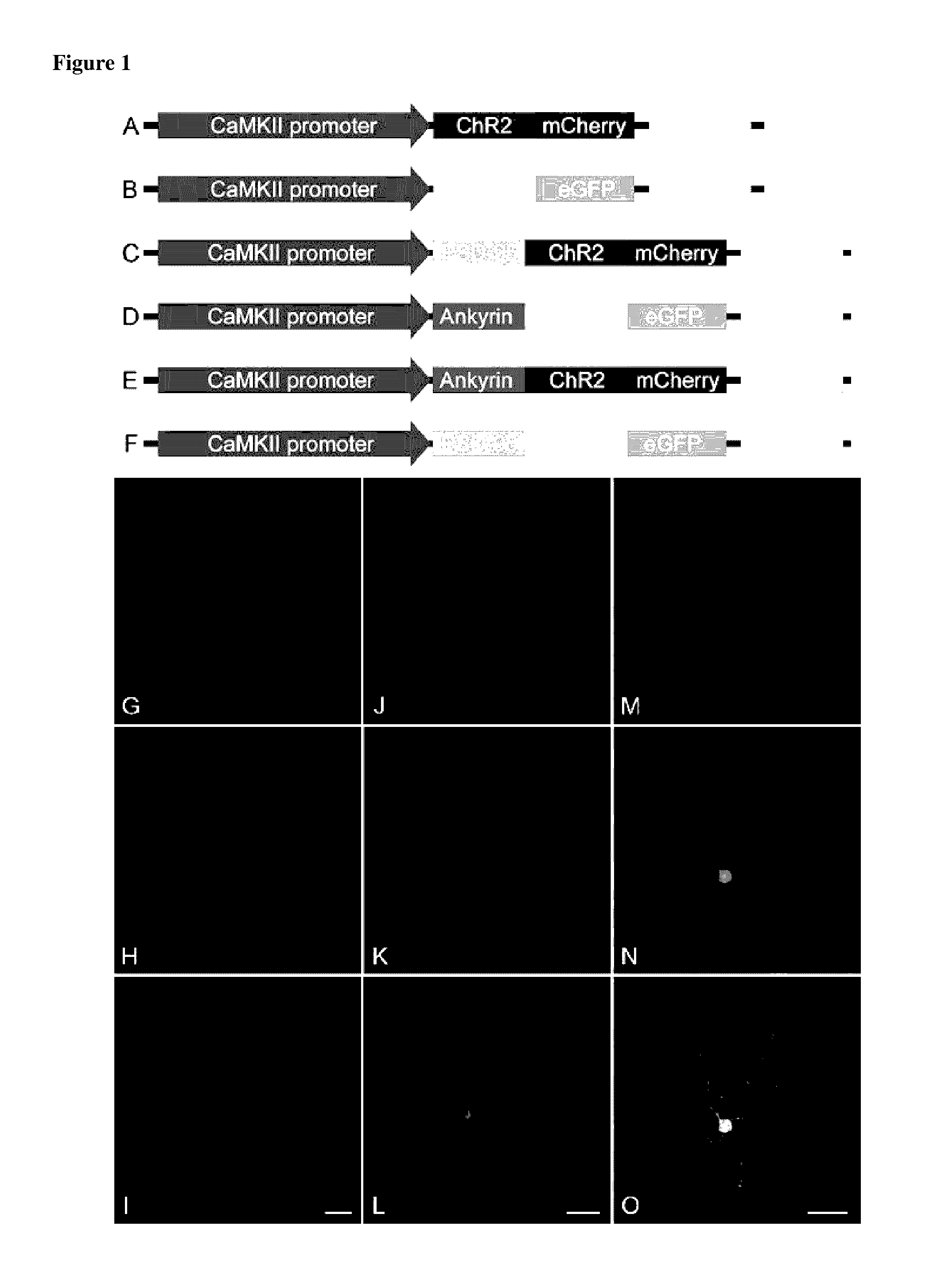
[0152]Proper synaptic development and function require precise localization of proteins to specialized subcellular and plasma membrane domains. Some classic examples are the accumulation of Na+ channels at nodes of Ranvier in association with the cytoskeletal protein ankyrin, the clustering of glycine receptors at inhibitory synapses in association with gephyrin, and accumulation of NMDA receptors and postsynaptic signaling cascades at excitatory synapses via postsynaptic density (PSD) proteins. Using these intrinsic localization mechanisms to create spatially distinct excitatory and inhibitory zones, we genetically targeted humanized channelrhodopsin-2 (hChR2), an excitatory cation channel, and enhanced halorhodopsin (eNpHR), an inhibitory chloride pump, to somatic or dendritic compartments. N-terminal fusions were constructed with these opsins and motifs derived from ankyrinG or postsynaptic density (PSD-95) proteins ...
example 2
HChR2 and eNpHR Behave Antagonistically at Normal Resting Potentials
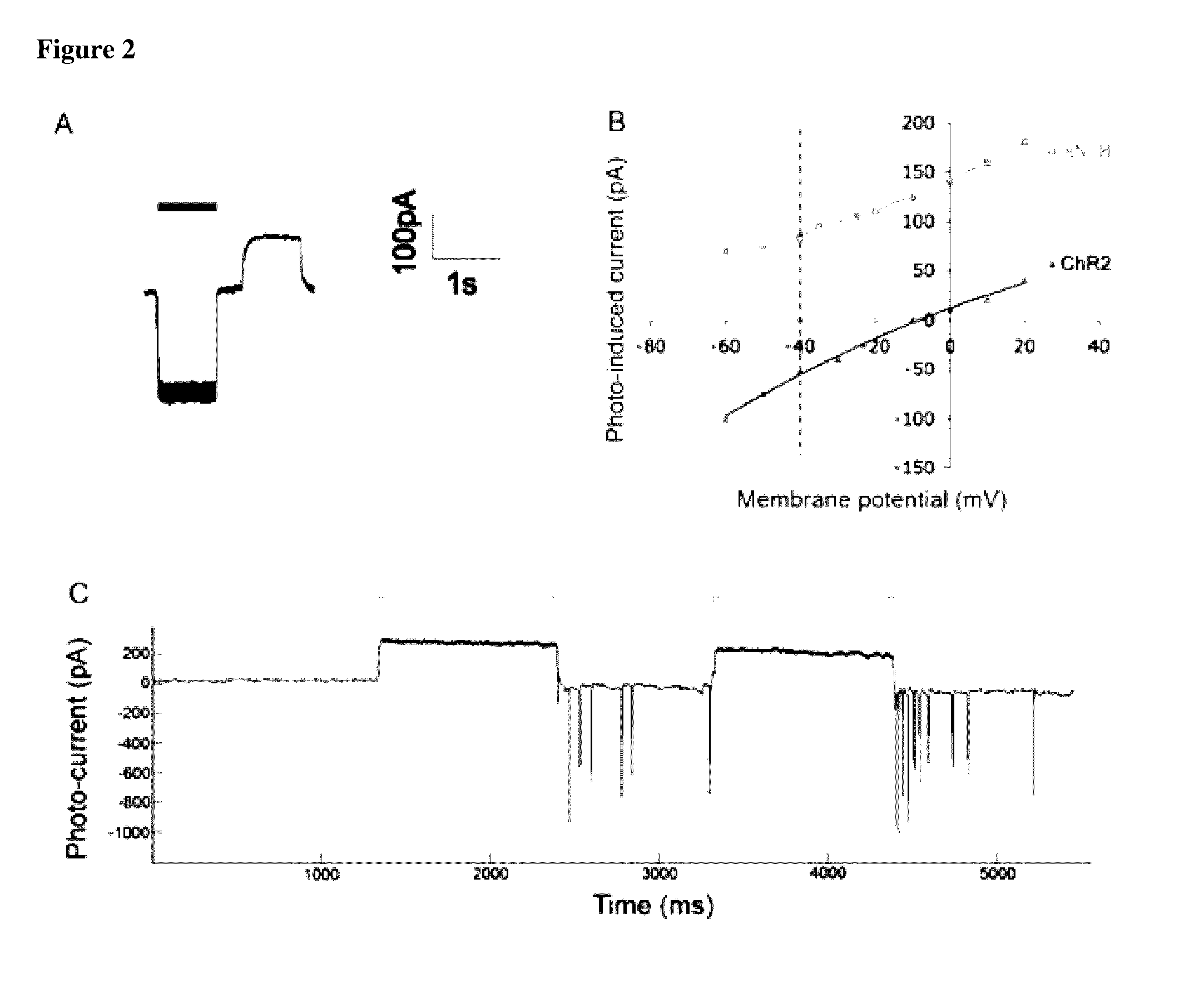
[0155]In order for genetically reconstructed center-surround antagonism to function effectively in a neuron, the opsins must generate opposing currents at physiologically-negative resting potentials and their chromatic sensitivity must be distinct. This antagonistic interaction was verified by whole-cell patch clamp recording from transfected ganglion cells in intact retina while perfusing a cocktail of CPP, 1-AP4, and CNQX to disrupt NMDA, metabotropic, and ionotropic glutamate receptor mediated currents derived from the photoreceptor to bipolar cell pathway. Following biolistic delivery (24-72 hrs) of PSD-95 and ankyrinG opsin fusions, we measured the electrophysiological properties of these transfected retinal ganglion cells in response to full field illumination. Currents were recorded from ganglion cells expressing both hChR2 and eNpHR while voltage clamped at negative resting potentials (−60 mV) during transie...
example 3
PSD95-hChR2Enables Robust Spiking in RGCs
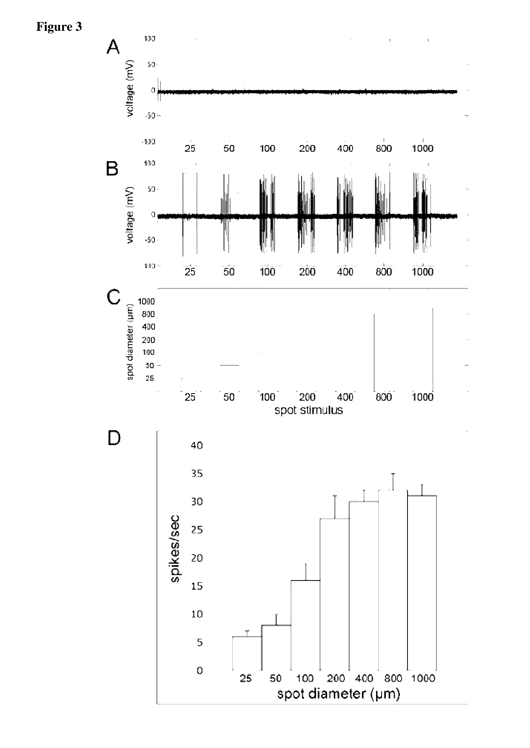
[0157]The PSD95-hChR2 fusion was effective at generating spikes in response to patterned blue illumination. Extracellular spike recordings were performed on ganglion cells expressing PSD95-hChR2 during illumination with white or blue spots of increasing diameter while the retina was perfused with synaptic transmission blockers (1-AP4, CPP, CNQX). White spots (0.012 mW / mm2) failed to evoke spike responses regardless of spot diameter (25-1000 μm). This illumination intensity is sufficient to drive photoreceptor-mediated spiking responses in light adapted rabbit retina in the absence of synaptic blockade (not shown), indicating that photoreceptor-mediated synaptic transmission was entirely blocked in the presence of the cocktail (FIG. 3A). In the same cell, blue spots (10 mW / mm2) elicited robust PSD95-hChR2 mediated spikes in response to spots ranging from 25-1000 μm (FIGS. 3B-3D). Spike frequency increased as the spot expanded to illuminate mor...
PUM
| Property | Measurement | Unit |
|---|---|---|
| whole cell voltage clamp recording | aaaaa | aaaaa |
| diameter | aaaaa | aaaaa |
| diameter | aaaaa | aaaaa |
Abstract
Description
Claims
Application Information
 - R&D
- Intellectual Property
- Life Sciences
- Materials
- Tech Scout
- Unparalleled Data Quality
- Higher Quality Content
- 60% Fewer Hallucinations
Browse by: Latest US Patents, China's latest patents, Technical Efficacy Thesaurus, Application Domain, Technology Topic, Popular Technical Reports.
© 2025 PatSnap. All rights reserved.Legal|Privacy policy|Modern Slavery Act Transparency Statement|Sitemap|About US| Contact US: help@patsnap.com


