Method and apparatus for automatic focusing of substrates in flourescence microscopy

a technology of flourescence microscopy and automatic focusing, which is applied in the direction of biochemistry apparatus and processes, color television, television systems, etc., can solve the problem of comparatively slow system

Active Publication Date: 2016-02-02
EUROIMMUN MEDIZINISCHE LABORDIAGNOSTIKA
View PDF5 Cites 4 Cited by
  • Summary
  • Abstract
  • Description
  • Claims
  • Application Information

AI Technical Summary

Benefits of technology

[0033]Thus, with the method described, it is possible in an advantageous manner to determine a focus level on the basis of a comparison of the recorded images in the evaluation unit. Those images recorded and likewise lying within the focus level are finally transferred to a device for preparing diagnostic findings. Preferably, this is a data processing unit, in particular ...

Problems solved by technology

Frequently, however, such systems are comparatively slow, since a multitu...

Method used


Image

  • Method and apparatus for automatic focusing of substrates in flourescence microscopy
  • Method and apparatus for automatic focusing of substrates in flourescence microscopy
  • Method and apparatus for automatic focusing of substrates in flourescence microscopy

Examples


Embodiment Construction

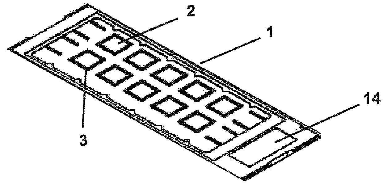
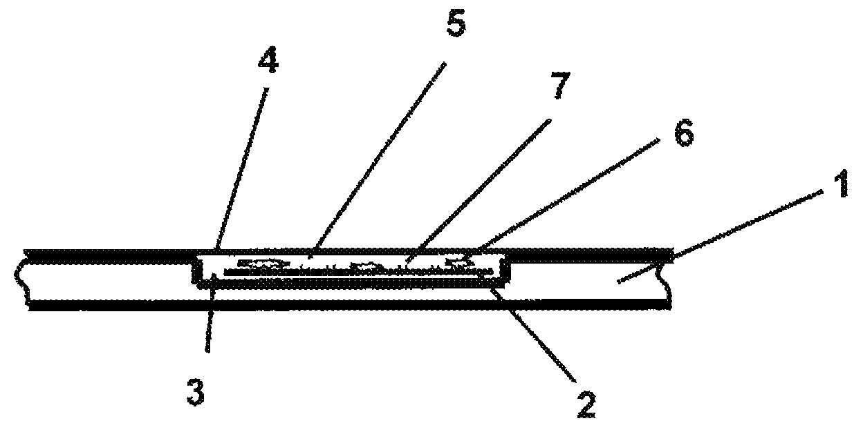
[0043]FIG. 1 first shows an object slide 1 with biochips 2 arranged thereon. In that, the object slide 1 has ten reaction fields 3, which compared to the remaining surface of the object slide 1 are executed as small depressions. The biochips 2 are arranged on the reaction fields 3. In principle, it is conceivable to provide one or even several biochips 2 on one reaction field 3. In this connection, it is self-evidently possible to adjust the size of the reaction field 3 in a suitable manner.

[0044]The biochips 2 are small slides with biological material, which were manufactured by coating of a standard cover slip with a tissue section and subsequent fragmentation of the cover slip. While a standard cover slip is an about 100 μm to 200 μm thin, rectangular or round glass platelet, which usually has an area of 18×18 mm2, biochips 2 are cover slip fragments coated with suitable biological material, which thus have a much smaller surface. Depending on the respective examination profile a...


PUM


Abstract

An apparatus for automated examination of biological material (6) includes a microscope (8) with a cross table (9) with the biological material (6) being arranged on the cross table (9) between an object slide (1) or an analysis plate and a cover (4); a light source (15, 16); an evaluation unit; a device for automatic focusing (11); an image recording unit (17) for recording an image of the biological material (6) enlarged via of an object lens (10) of the microscope (8) and for transferring the image to the evaluation unit; and a biochip (2). Labeling (7) is provided on a surface of the biochip (2). Focusing is executable by targeted movement of the cross table (9) based on detection of the labeling (7), and a target interval is determinable by determination of a location and/or a position of the labeling, within which focusing of the biological material takes place.

Description

BACKGROUND OF THE INVENTION[0001]The instant application should be granted the priority dates of Aug. 23, 2010, the filing date of the corresponding German patent application 102010035104.0, as well as Aug. 23, 2011, the filing date of the International patent application PCT / EP2011 / 004227.[0002]The invention relates to a method as well as an apparatus for automated examination of biological material with a microscope, on the cross table of which the biological material is arranged between an object slide or an analysis plate and at least one cover, with at least one light source for exposure of the biological material as well as with an image recording unit recording at least one image of the biological material enlarged by means of the object lens of the microscope and transferring it to an evaluation unit. The technical solution described provides labeling, which is detectable by a device for automatic focusing of the biological material, wherein the focusing can be executed by t...

Claims


Application Information

Patent Timeline
IPC IPC(8): G06K9/00G02B21/34G02B21/00G02B7/36G02B21/24G01N21/64C12Q1/00
CPCG02B21/34G02B7/36G02B21/0004G02B21/241G01N21/6458
Inventor MORRIN, MARKUS
Owner EUROIMMUN MEDIZINISCHE LABORDIAGNOSTIKA
Why Eureka
Try Eureka