
Method and system for fluorescent imaging with background surgical image composed of selective illumination spectra

a fluorescent imaging and selective illumination technology, applied in the field of endoscopic imaging, can solve the problems of difficult identification of certain tissue types, too small picture in the pip display, and achieve the effect of reducing the memory and processing requirements of the system

Active Publication Date: 2015-12-15
INTUITIVE SURGICAL OPERATIONS INC
View PDF32 Cites 5 Cited by
  • Summary
  • Abstract
  • Description
  • Claims
  • Application Information

AI Technical Summary

Benefits of technology

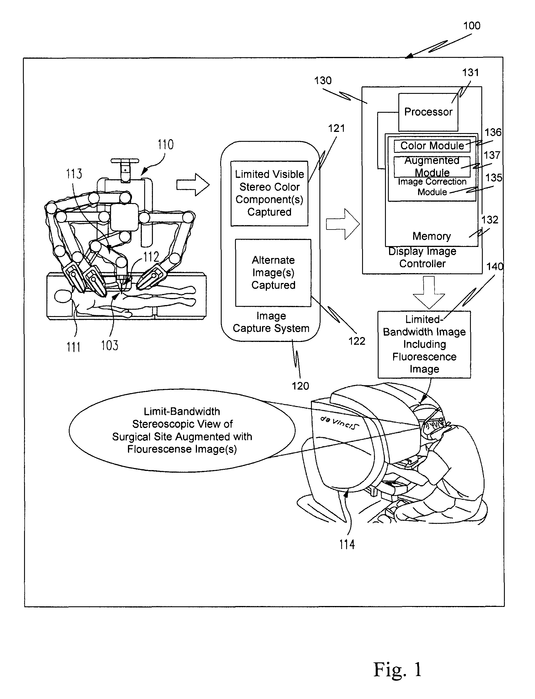
This patent describes a method for providing real-time images to a surgeon during minimally invasive surgeries. The method involves using a special light to highlight important areas of the surgical site, which are then combined with a normal image to create a single display for the surgeon. This is done without introducing any delay or adding extra processing requirements, resulting in a more efficient and effective display. The method also reduces the system's memory and processing requirements compared to other methods that use time slicing. Overall, this technology enables better visual guidance for surgeons during surgeries, leading to improved outcomes for patients.

Problems solved by technology

In a typical surgical field, however, certain tissue types are difficult to identify, or tissue of interest may be at least partially obscured by other tissue.
However, the smaller picture in a PIP display may be too small to show a desirable level of detailed information.
Moreover with separate image frames, a surgeon mentally fuses the two separate images or imaging modalities together, which can be fatiguing.

Method used


Image

  • Method and system for fluorescent imaging with background surgical image composed of selective illumination spectra
  • Method and system for fluorescent imaging with background surgical image composed of selective illumination spectra
  • Method and system for fluorescent imaging with background surgical image composed of selective illumination spectra

Examples


Embodiment Construction

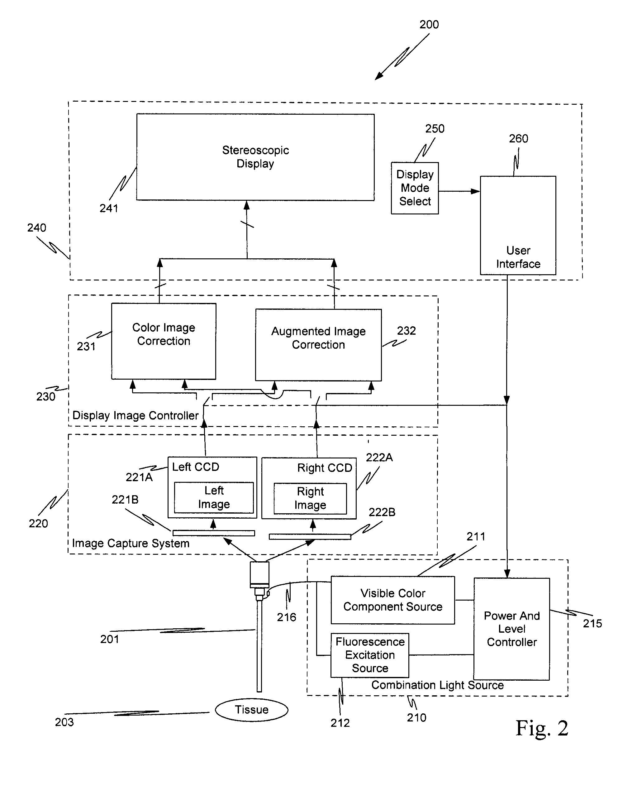
[0040]As used herein, electronic stereoscopic imaging includes the use of two imaging channels (i.e., channels for left and right images).

[0041]As used herein, a stereoscopic optical path includes two channels in an endoscope for transporting light from tissue (e.g., channels for left and right images). The light transported in each channel represents a different view of the tissue. The light can include one or more images. Without loss of generality or applicability, the aspects described more completely below also could be used in the context of a field sequential stereo acquisition system and / or a field sequential display system.

[0042]As used herein, an illumination path includes a path in an endoscope providing illumination to tissue.

[0043]As used herein, images captured in the visible electromagnetic radiation spectrum are referred to as acquired visible images.

[0044]As used herein, white light is visible white light that is made up of three (or more) visible color components, ...


PUM


Abstract

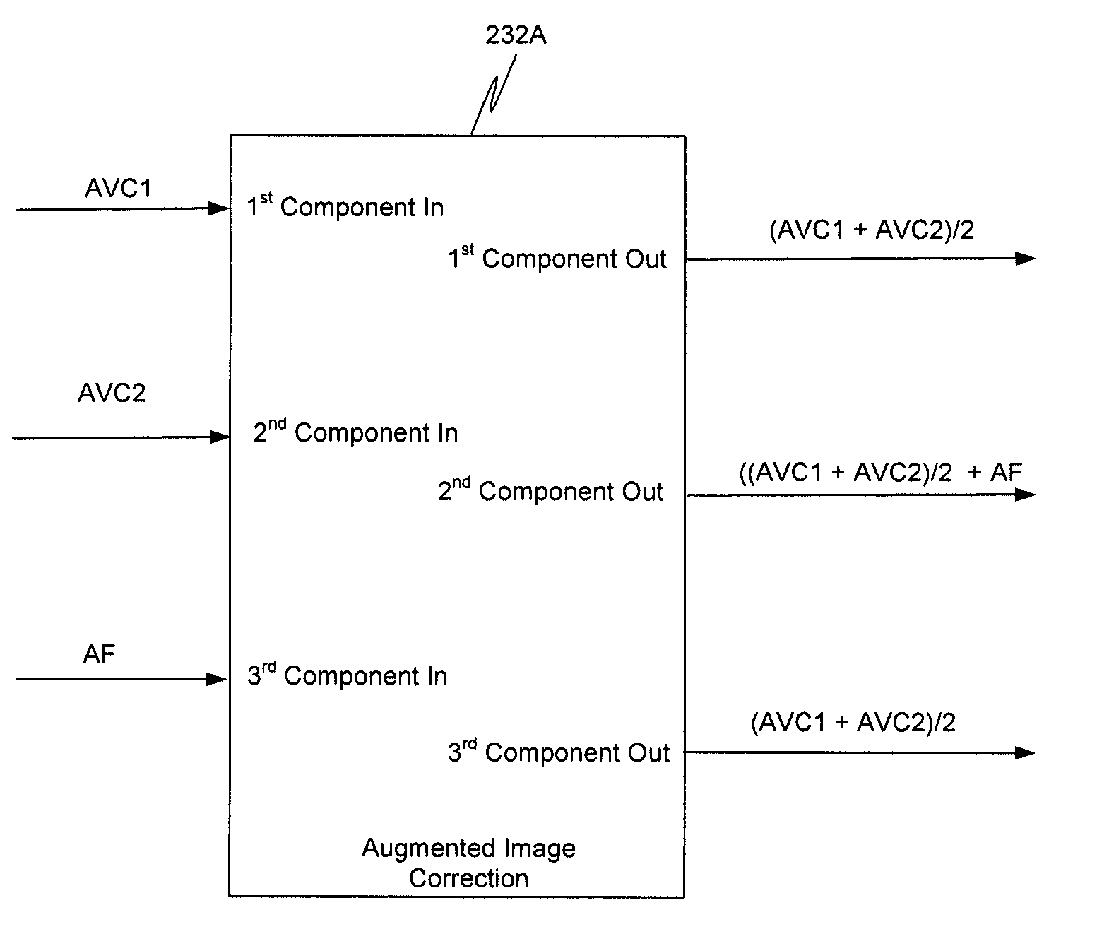
A surgical site is simultaneously illuminated by less than all the visible color components that make up visible white light, and a fluorescence excitation illumination component by an illuminator in a minimally invasive surgical system. An image capture system acquires an image for each of the visible color components illuminating the surgical site and a fluorescence image, which is excited by the fluorescence excitation component from the illuminator. The minimally invasive surgical system uses the acquired images to generate a background black and white image of the surgical site. The acquired fluorescence image is superimposed on the background black and white image, and is highlighted in a selected color, e.g., green. The background black and white image with the superimposed highlighted fluorescence image is displayed for a user of the system. The highlighted fluorescence image identifies tissue of clinical interest.

Description

RELATED APPLICATION[0001]This application claims priority to and the benefit of:[0002]U.S. Provisional Application No. 61 / 361,260 filed Jul. 2, 2010 entitled “METHOD AND SYSTEM FOR FLUORESCENT IMAGING WITH BACKGROUND SURGICAL IMAGE COMPOSED OF SELECTIVE ILLUMINATION SPECTRA,” naming as inventors, Ian McDowall and Christopher J. Hasser, which is incorporated herein by reference in its entirety.BACKGROUND[0003]1. Field of Invention[0004]Aspects of this invention are related to endoscopic imaging, and are more particularly related to blending visible and alternate images so as to provide an enhanced real-time video display for a surgeon.[0005]2. Related Art[0006]The da Vinci® Surgical System, commercialized by Intuitive Surgical, Inc., Sunnyvale, Calif., is a minimally invasive teleoperated surgical system that offers patients many benefits, such as reduced trauma to the body, faster recovery and shorter hospital stay. One key component of the da Vinci® Surgical System is a capability ...

Claims


Application Information

Patent Timeline
Patent Type & Authority Patents(United States)
IPC IPC(8): A61B1/06A61B1/04A61B1/00A61B5/00A61B34/35A61B90/00
CPCA61B1/0638A61B1/00186A61B1/00193A61B1/043A61B5/0071A61B5/0084A61B1/063A61B1/0684A61B5/7425A61B1/046
Inventor MCDOWALL, IAN E.HASSER, CHRISTOPHER J.
Owner INTUITIVE SURGICAL OPERATIONS INC
Learn More