
Affinity-based detection of biological targets

a technology of biological targets and affinity-based detection, which is applied in the field of biological detection, can solve the problems of increasing the complexity and potential for non-specific or cross-reactive binding, increasing the cost and time-consuming of large-scale production of monoclonals, and antibody-based assays may never be stable enough for long-term sensor applications

Active Publication Date: 2014-02-25
UNITED STATES OF AMERICA
View PDF12 Cites 2 Cited by
  • Summary
  • Abstract
  • Description
  • Claims
  • Application Information

AI Technical Summary

Benefits of technology

The invention is a method and device for identifying biological targets using capture species such as antimicrobial peptides, cytotoxic peptides, antibiotics, and combinations thereof. The method involves detecting which capture species have bound biological targets to create a binding pattern, which is then used to identify the target. The device includes one or more substrates with a plurality of independent capture species bound to them, which can include antimicrobial peptides, cytotoxic peptides, antibiotics, and combinations thereof. These capture species can also have multiple specificities, meaning they can bind to multiple targets. The technical effect of this invention is a more efficient and effective method for identifying biological targets and their related molecular interactions.

Problems solved by technology

However, assays utilizing antibodies for specific recognition of target analytes have a number of problems that may significantly limit their widespread use in the field: 1) many antibodies are sensitive to environmental temperatures and must be stored frozen, refrigerated, or lyophilized for retention of optimal activity; 2) at least one antibody or set of antibodies is required for each target of interest in multiplexed assays, increasing the complexity and potential for non-specific or cross-reactive binding; 3) specificity and sensitivity of antibody-based recognition may, in some cases, be mutually exclusive; 4) target-specific antibodies may not be available due to the non-antigenic nature of the analyte; and 5) although monoclonal antibodies are, by their very nature, more consistent than polyclonal antibodies, development and large-scale production of monoclonals is expensive and time-consuming.
However, antibody-based assays may never be stable enough for long-term sensor applications; such stability is critical for fielding sentry-type systems and for non-laboratory use.

Method used


Image

  • Affinity-based detection of biological targets
  • Affinity-based detection of biological targets
  • Affinity-based detection of biological targets

Examples


example 1

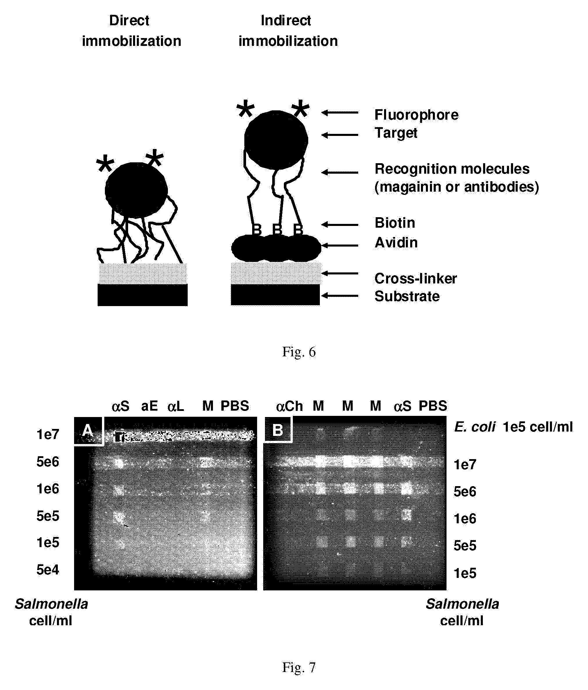
[0074]Array Biosensor—Immobilization of peptides and antibodies—FIG. 6 schematically illustrates immobilization of AMPs on a glass slide followed by binding of a fluorescent target. The direct method has attachment of recognition molecules to the substrate through cross-linking chemistry with GMBS. Indirect immobilization has attachment of recognition molecules to the substrate through an avidin-biotin interaction. The immobilization methodology utilizes sequential incubations with a thiol-terminated silane, the heterobifunctional crosslinker N-[γ-maleimidobutyryloxy]-succinimide ester (GMBS), and a protein or peptide containing one or more primary amines. Briefly, standard soda-lime microscope slides (Daigger, Vernon Hills, Ill.) cleaned with 10% KOH (w / v) in methanol were treated for 1 hour, under nitrogen, with a 2% solution of 3-mercaptopropyl trimethoxysilane in toluene. The slides were then rinsed with toluene, dried under nitrogen, and incubated for 30 minutes in 1 mM GMBS cr...

example 2

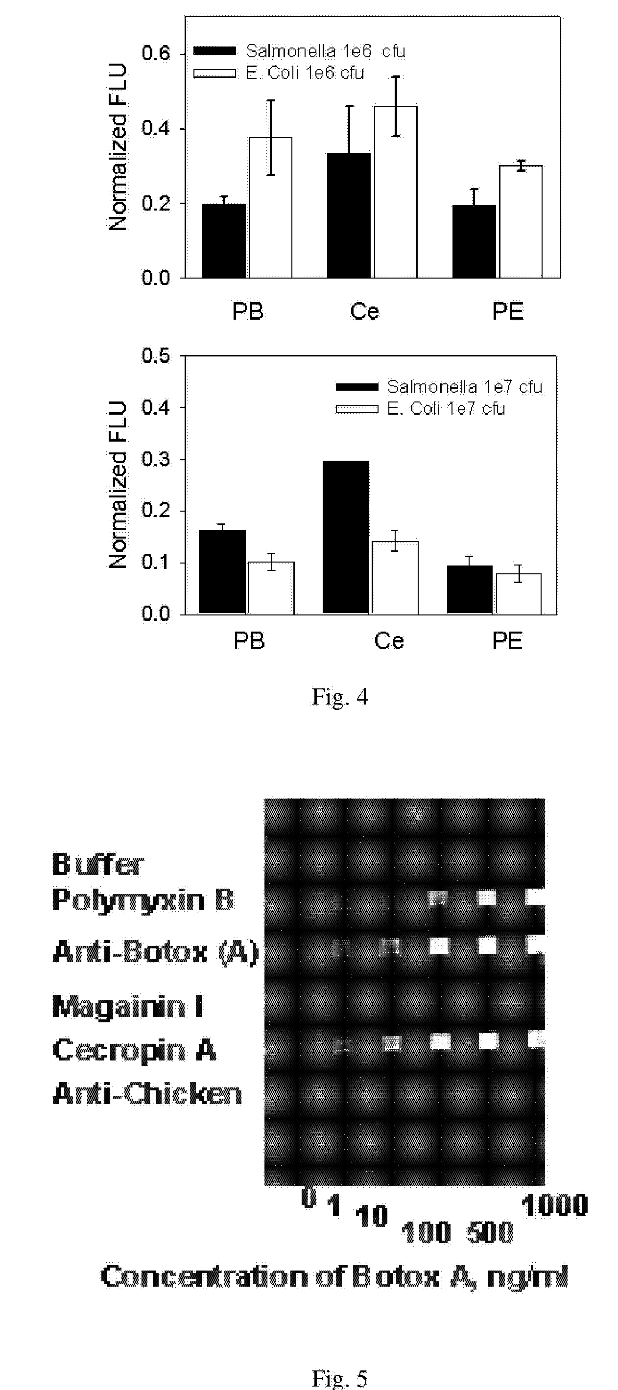
[0098]Luminex—“Sandwich”-type Luminex assays had been developed, using antibodies for target capture (initial recognition) and AMPs for detection of bound targets (FIG. 10, format A). Development of AMP-capture assays for Luminex (FIG. 10, format B) has begun; these results from Luminex should be more comparable to those from the Array Biosensor for which AMP-capture assays have been developed. To date, this system has successfully shown capture of E. coli, Salmonella, and bot toxoid A using AMP-derivatized Luminex beads (FIG. 11). Although LODs for E. coli and Salmonella were the same as those obtained in antibody-based Luminex assays (102-103 on bactenecin and 105 cfu / ml on PMB, respectively), bot toxoid A could be detected only at extremely high concentrations (1 μg / ml); these latter results contrast greatly with results obtained using the Array Biosensor. Neither Y pestis nor Coxiella could be detected at the concentrations tested on Luminex using AMP-coated Luminex beads for ca...

example 3

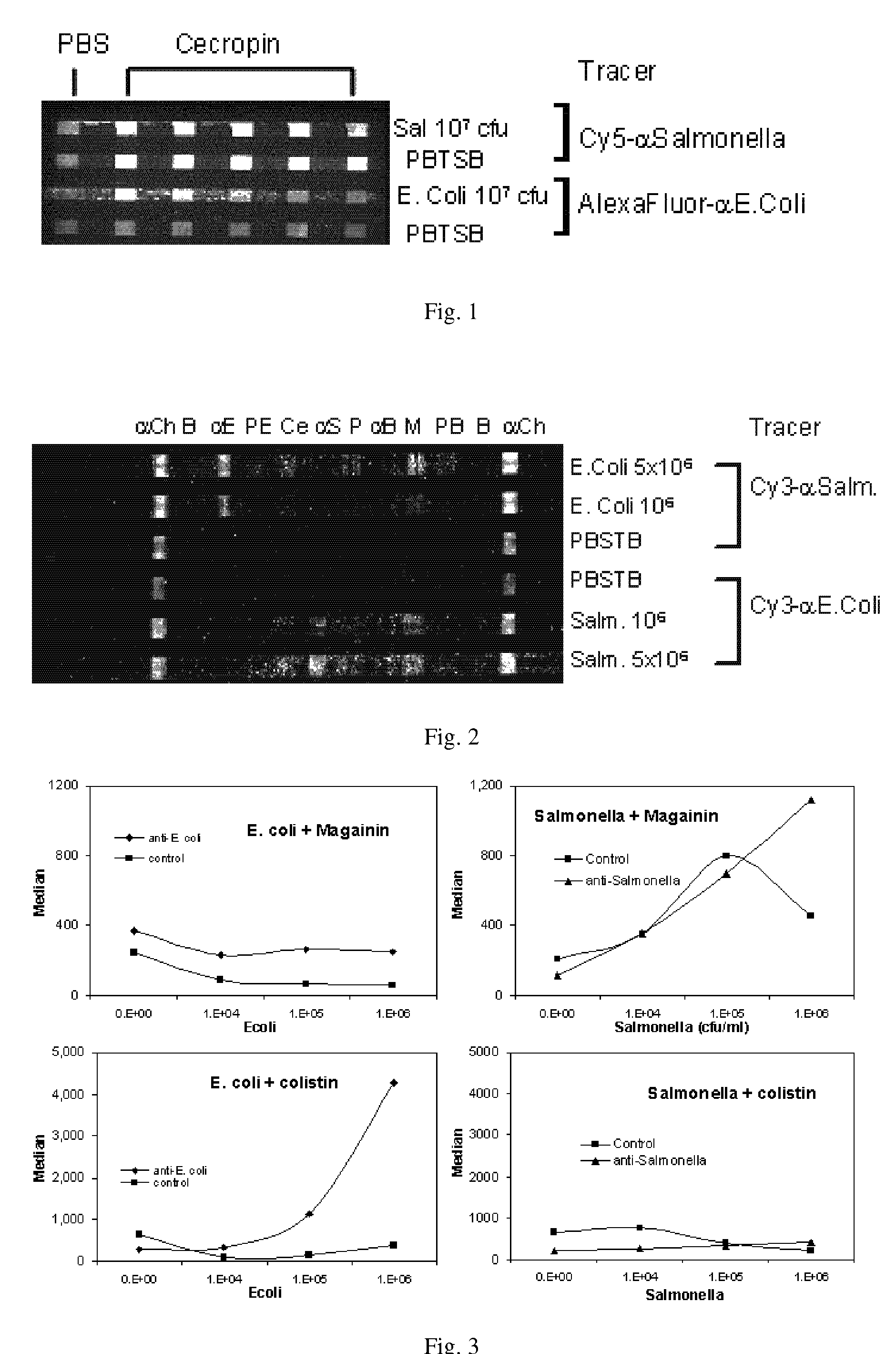
[0100]Binding patterns—Above, differences in binding between E. coli and Salmonella, two very closely related species, using AMP tracer beads in Luminex assays are demonstrated. This was extended to include additional species on Luminex using AMP tracers (FIG. 12) and as well as AMP-Lx beads for capture (FIG. 13). Similar results have been observed in the Array Biosensor (FIG. 14). Several particularly noteworthy observations have been made with regard to binding patterns. Bot toxoids A and B, antigenically similar proteins, bind to different AMPs. The binding affinities for melittin in the assays run contrary to the hypothesis that inhibition of bot toxins by various AMPs is most likely caused by AMP binding to active sites. Melittin contains the KR sequence required for cleavage by bot toxin A peptidase activity, but is bound preferentially by toxoid B; conversely, cecropin A is bound preferentially by toxoid A, but does not contain any of the sequences required for cleavage by to...


PUM

PropertyMeasurementUnit
pHaaaaaaaaaa
concentrationaaaaaaaaaa
pHaaaaaaaaaa

Abstract

A method of biochemical identification by: providing a plurality of capture species bound to one or more substrates and suspected of having one or more biological targets affinity bound to at least one capture species; detecting which capture species contain bound biological targets to generate a binding pattern; and identifying the biological target based on the binding pattern. The capture species are independently selected from the group consisting of antimicrobial peptides, cytotoxic peptides, antibiotics, and combinations thereof. A device having the capture species bound to the substrates. At least two of the capture species are capable of multi-specific binding to one or more biological targets and may have overlapping but not identical affinity properties.

Description

CROSS REFERENCE TO RELATED APPLICATIONS[0001]This application claims priority to U.S. Provisional Patent Application No. 60 / 690,046, filed on Jun. 10, 2005, incorporated herein by reference. All other referenced patent documents and publications are incorporated herein by reference.FIELD OF THE INVENTION[0002]The invention is generally related to biological detection.DESCRIPTION OF RELATED ART[0003]With the notable exception of glucose sensors, the vast majority of rapid detection / measurement systems use antibodies for recognition, identification, and quantification of biological targets. Antibody-based detection techniques are powerful, versatile tools for various molecular and cellular analyses, environmental monitoring, and clinical diagnostics. This power originates from the specificity of the antibodies for their particular antigenic sites.[0004]Antibody-based recognition of targets is the basis for detection in many optical and electrochemical biosensors (e.g., interferometers...

Claims


Application Information

Patent Timeline
Patent Type & Authority Patents(United States)
IPC IPC(8): G01N33/53
CPCG01N33/56916G01N33/554G01N33/54366G01N33/5308G01N33/56983G01N33/56911
Inventor KULAGINA, NADEZHDA VTAITT, CHRIS ROWEANDERSON, GEORGE PLIGLER, FRANCES S
Owner UNITED STATES OF AMERICA
Learn More