
Method for imaging in magnetic resonance tomography with spectral fat saturation or spectral water excitation

a magnetic resonance tomography and fat saturation technology, applied in the field of resonance tomography (mrt), can solve the problems of low overall signal yield, low signal to noise ratio with low anatomical detail of the image, fat signal often reduces the distinguishability of details in mr images, etc., and achieves significant improvement in image quality

Inactive Publication Date: 2013-03-12
SIEMENS HEALTHCARE GMBH
View PDF4 Cites 3 Cited by
  • Summary
  • Abstract
  • Description
  • Claims
  • Application Information

AI Technical Summary

Benefits of technology

This invention is a way to get magnetic resonance images that have better quality. It uses a technique called spectral saturation or excitation to improve the image.

Problems solved by technology

The presence of both fat and water tissue in a patient constitutes one challenge in magnetic resonance imaging.
Additionally, the fat signal often reduces the distinguishability of details in MR images both in T1-imaging and in T2 imaging because of its high signal contribution to the overall MR signal.
Along with the relatively long acquisition time the disadvantage of STIR is among other things the relatively low overall signal yield (poor signal to noise ratio with low anatomical detail of the image).
Another disadvantage is the fact that STIR cannot be used for contrast agent based MRT measurements.
However, the 2-point Dixon method fails in the case of voxels, in which the signals of fat and water in cancel each other out, which thus contain approximately equal amounts of water and fat.
However in the regions of the cervical vertebral column that have already been mentioned as well as in the knee, the many different tissue volumes change over a few centimeters so starkly that, due to the changing susceptibility values associated with this (tissue to air approximately 6 ppm), the B0 field variation with respect to the overall image cannot be compensated in spite of the shimming.
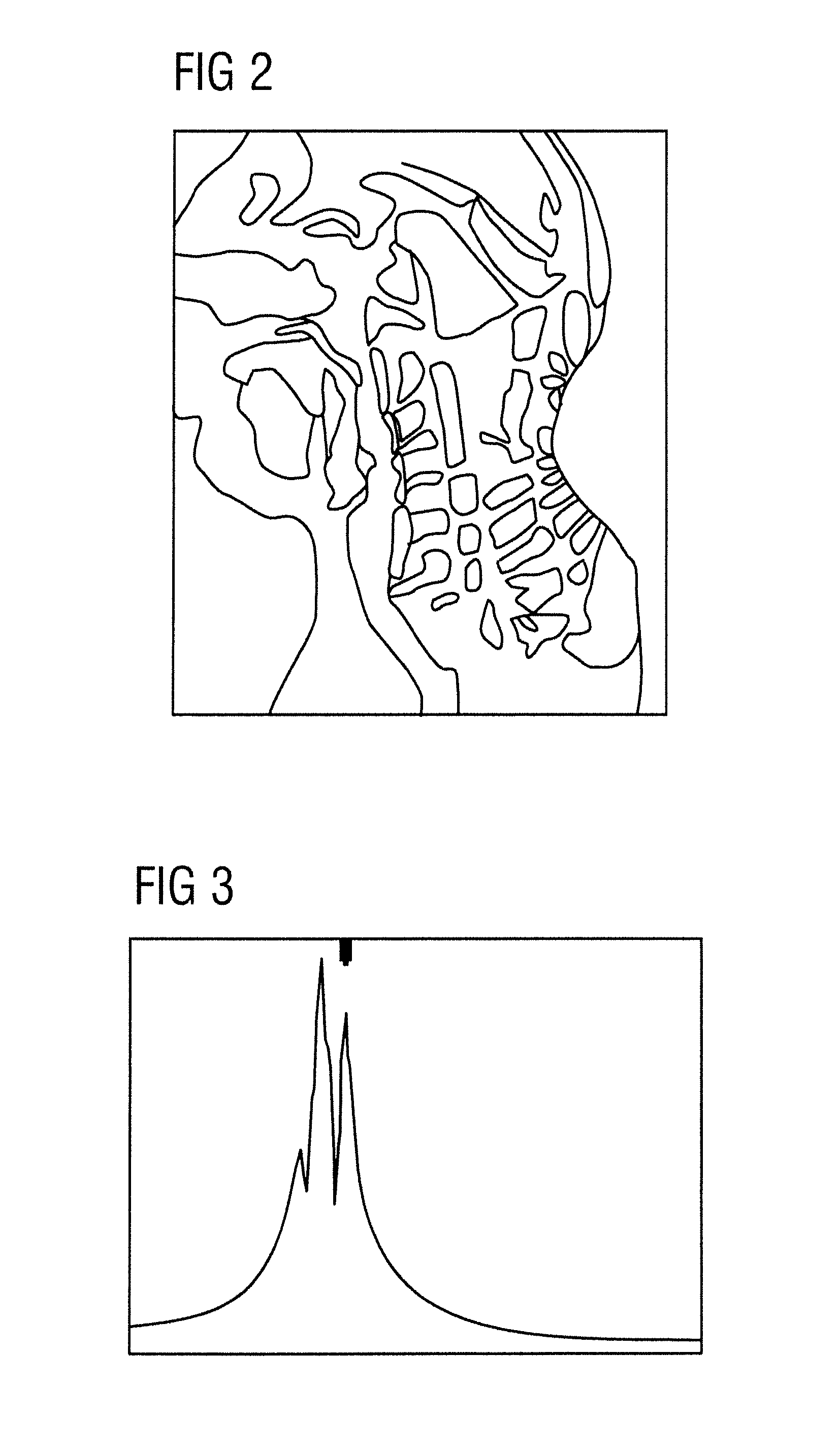
Within the scope of the necessary frequency adjustment (also after preceding shimming) the resonant frequency can then only be set with insufficient precision from the measured superimposed lines of the spectrum, which in individual critical partial images leads to undesirable saturation effects.

Method used


Image

  • Method for imaging in magnetic resonance tomography with spectral fat saturation or spectral water excitation
  • Method for imaging in magnetic resonance tomography with spectral fat saturation or spectral water excitation
  • Method for imaging in magnetic resonance tomography with spectral fat saturation or spectral water excitation

Examples


Embodiment Construction

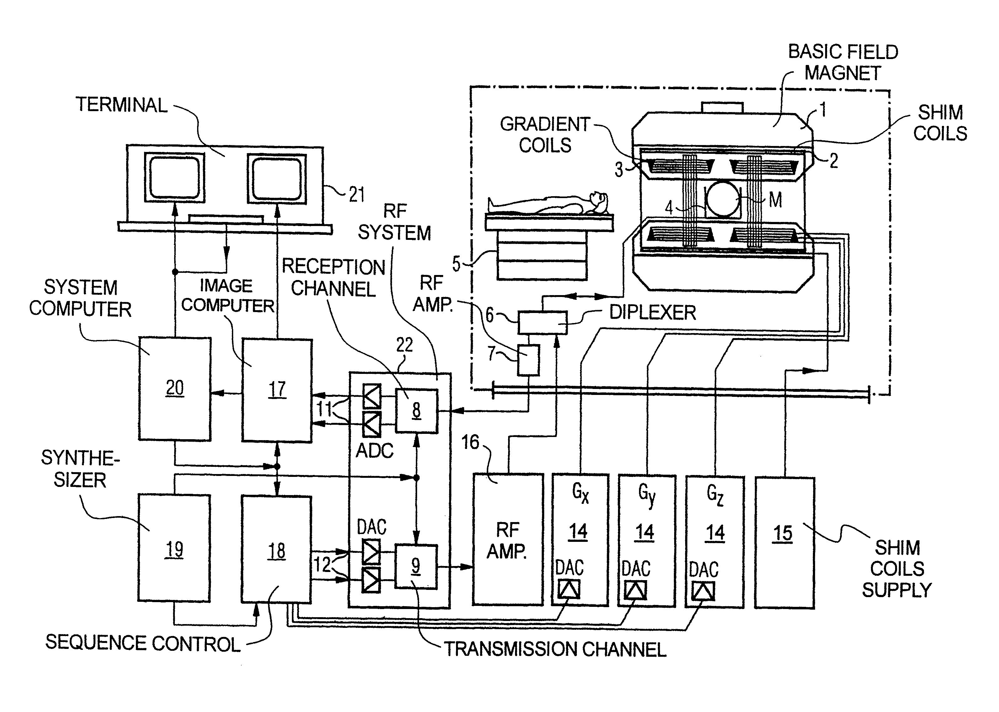
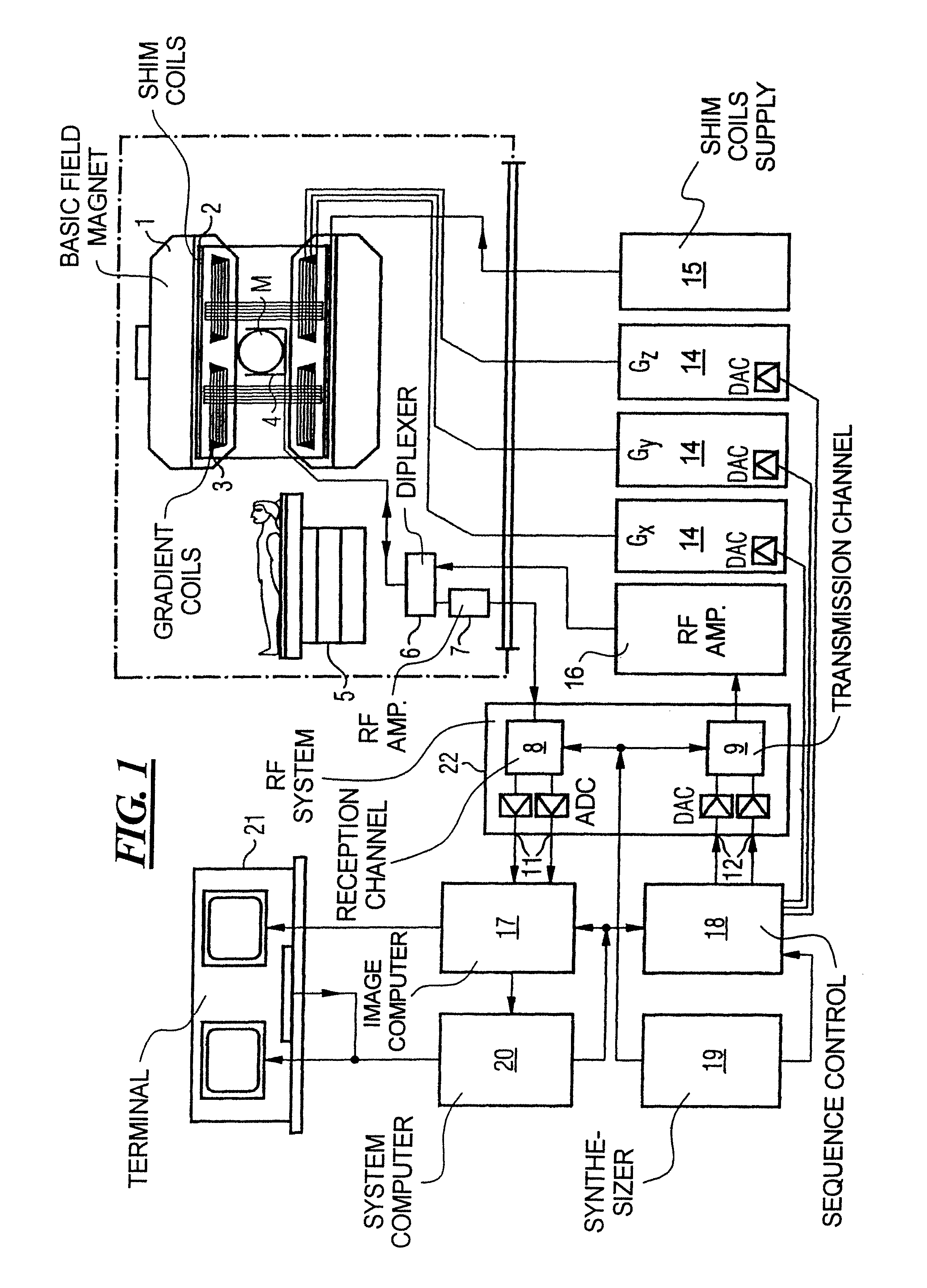
[0043]FIG. 1 schematically shows a magnetic resonance imaging (tomography) device for the generation of a magnetic resonance image of an object in accordance with the present invention. The basic components of the tomography device correspond to those of a conventional tomography device, with differences discussed below. A basic field magnet 1 generates a temporally constant strong magnetic field for polarization or alignment of the nuclear spin in the region under examination of an object, e.g., a part of a human body to be examined. The high homogeneity of the basic field magnet required for the magnetic resonance data acquisition is defined in a measurement volume V, in which the parts of the human body to be examined are inserted. For support of the homogeneity requirements and in particular for the elimination of temporally invariable influences, so-called shims made of ferromagnetic material are mounted at suitable locations. Temporally variable influences are eliminated by sh...


PUM


Abstract

A magnetic resonance tomography (MRT) method with spectral fat saturation or spectral water excitation in a tissue region that is to be represented of a patient who is to be examined, includes the following steps: (Step 1) frequency adjustment measurement of a region of a patient that is to be represented with a selected first partial coil of the MRT system, (Step 2) precise determination of the resonance frequency of water with the aid of the spectrum obtained in Step 1 exhibiting the resonance frequencies of fat and water, (Step 3) repetition of Steps 1 and 2 with at least one additionally selected second partial coil of the MRT system adjacent to the first partial coil, (Step 4) measuring of a k space data record with a partial coil or a partial coil combination on the basis of the water resonance frequency assigned to these partial coils, (Step 5) repetition of Step 4 with other partial coils or other partial coil combinations until the entire tissue region to be represented has been measured, (Step 6) combining of the measuring results obtained in Steps 4 and 5, and (Step 7) representing of the results obtained in Step 6 in the image space in the form of an overall image of the tissue region to be represented.

Description

BACKGROUND OF THE INVENTION[0001]1. Field of the Invention[0002]The present invention relates in general to resonance tomography (MRT), as IS applicable in medicine for the examination of patients. The present invention relates in particular to an MRT method in which the image quality is improved in the case of images with spectral saturation or excitation.[0003]2. Description of the Prior Art[0004]Magnetic resonance tomography is a tomographic imaging modality for medical diagnostics, which is characterized first and foremost by a high contrast resolution capability. On the basis of an outstanding ability to represent soft tissue, magnetic resonance tomography has developed to a method far superior to X-ray computed tomography. Magnetic resonance tomography today is based on the application of spin echo and gradient echo sequences, which in the case of measuring times of the order of minutes makes an excellent image quality possible.[0005]The presence of both fat and water tissue i...

Claims


Application Information

Patent Timeline
Patent Type & Authority Patents(United States)
IPC IPC(8): G01V3/00
CPCG01R33/3415G01R33/4828G01R33/5607G01R33/56527
Inventor DEIMLING, MICHAEL
Owner SIEMENS HEALTHCARE GMBH
Learn More