Activation of bone and cartilage formation
a technology of activation and cartilage, applied in the field of tissue treatment systems, can solve the problems of reducing the rate of wound healing, affecting the effect of healing, and affecting the quality of life of patients, and achieve the effect of reducing pressur
- Summary
- Abstract
- Description
- Claims
- Application Information
AI Technical Summary
Benefits of technology
Problems solved by technology
Method used
Image
Examples
example 1
Stimulation of Osteogenic Activity in the Dura Mater with Reduced Pressure
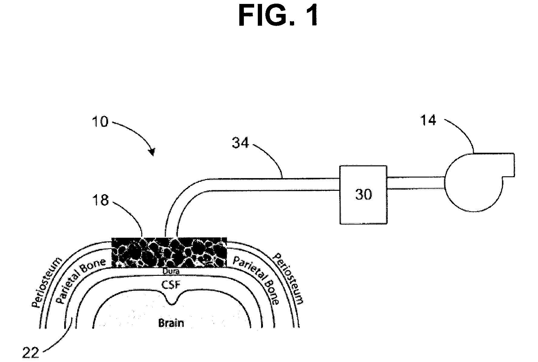
[0055]Referring to FIG. 2, a cranial defect study was performed to evaluate the effects of applying reduced pressure to intact dura mater through a scaffold. Critical size defects were made in the cranium of a rabbit with dura mater left intact. A scaffold similar to that shown in FIG. 1 (i.e. manifold and / or scaffold 18) was placed in contact with the dura mater. Several tests were conducted that varied the amount of time over which reduced pressure was applied. A control test was run in which no reduced pressure was supplied to the scaffold placed in contact with the dura mater. Samples of the cranium and scaffold were examined following 12 weeks of in-life healing for each particular defect (inclusive of the amount of time over which reduced pressure was applied). The measured values included the amount of new bone area observed, the percentage of quantitative bridge assessment, the percentage of total scaf...
example 2
Stimulation of a Periosteum Osteogenic Response by Foam Manifolds and Reduced Pressure
[0060]A foam manifold and reduced pressure were evaluated for their ability to induce the periosteum to synthesize new bone. The intact, undamaged cranial periosteum of rabbits was exposed. A GranuFoam® (KCI Licensing, Inc., San Antonio Tex.) foam dressing was applied to the bone. In some treatments, the foam-covered bone was also subjected to reduced pressure. After treatment, the animals were sacrificed and the treated bone was subjected to paraffin embedding, sectioning and staining to evaluate the effect of the treatment on new bone formation.
[0061]FIG. 5 shows a naïve, undamaged periosteum in a rabbit. The arrow denotes the location of the thin layer of the periosteum. The periosteum is thin and unremarkable. By contrast, FIG. 6 shows induction of granulation tissue overlying the periosteum where the foam was maintained in contact with the bony surface for 6 days without reduced pressure. Gran...
example 3
Induction of Cartilage Tissue Formation
[0063]Cartilage formation was observed in response to the application of reduced pressure therapy to the surface of intact cranial periosteal membranes. These observations are of significance in that cartilage formation in response to a therapy is unique and of great interest in the field of tissue engineering. These formations were observed in the absence of scaffold materials and only with the application of reduced pressure. No cartilage formation was observed in controls.
[0064]Cartilage degeneration caused by congenital abnormalities or disease and trauma is of great clinical consequence. Because of the lack of blood supply and subsequent wound-healing response, damage to cartilage generally results in an incomplete repair by the body. Full-thickness articular cartilage damage, or osteochondral lesions, allow for the normal inflammatory response, but result in inferior fibrocartilage formation. Surgical intervention is often the only option...
PUM
 Abstract
Description
Claims
Application Information
 - R&D
- Intellectual Property
- Life Sciences
- Materials
- Tech Scout
- Unparalleled Data Quality
- Higher Quality Content
- 60% Fewer Hallucinations
Browse by: Latest US Patents, China's latest patents, Technical Efficacy Thesaurus, Application Domain, Technology Topic, Popular Technical Reports.
© 2025 PatSnap. All rights reserved.Legal|Privacy policy|Modern Slavery Act Transparency Statement|Sitemap|About US| Contact US: help@patsnap.com


