
System and method for three-dimensional mapping of electrophysiology information

a three-dimensional mapping and electrophysiology information technology, applied in the field of electrophysiology equipment, can solve the problems of only sparse innervation of the ventricular myocardium by the efferent vagal nerve, and the rhythmic contraction of the heart muscl

Active Publication Date: 2011-10-18
ST JUDE MEDICAL ATRIAL FIBRILLATION DIV
View PDF72 Cites 69 Cited by
  • Summary
  • Abstract
  • Description
  • Claims
  • Application Information

AI Technical Summary

Benefits of technology

The present invention expands the capabilities of cardiac electrophysiology mapping systems to provide additional diagnostic data using both the time domain and frequency domain representations of electrophysiology data. This creates a three-dimensional map of the electrical activity and related information. The system can create maps that show the time difference between action potentials, peak-to-peak voltage, peak negative voltage, complex fractionated electrogram information, dominant frequency, maximum peak amplitude, ratio of energy in different frequency bands, low-frequency or high-frequency passband, frequency with maximum energy, number of peaks in a passband, energy, power, and area in each peak, ratio of energy and area in each peak to another passband, and width of each peak in a spectrum. The system can also update the colors, shades of colors, and grayscales of the parameters on the three-dimensional model. Overall, this invention allows for more accurate and comprehensive mapping of cardiac electrical activity.

Problems solved by technology

First, they generate periodic impulses that cause rhythmical contraction of the heart muscle.
Second, they conduct the periodic impulses rapidly throughout the heart.
However, efferent vagal nerves only sparsely innervate the ventricular myocardium.
Altered impulse conduction commonly results in reentry, which can lead to tachyarrhythmias.

Method used


Image

  • System and method for three-dimensional mapping of electrophysiology information
  • System and method for three-dimensional mapping of electrophysiology information
  • System and method for three-dimensional mapping of electrophysiology information

Examples


Embodiment Construction

[0025]System Level Overview and Basic Location Methodology

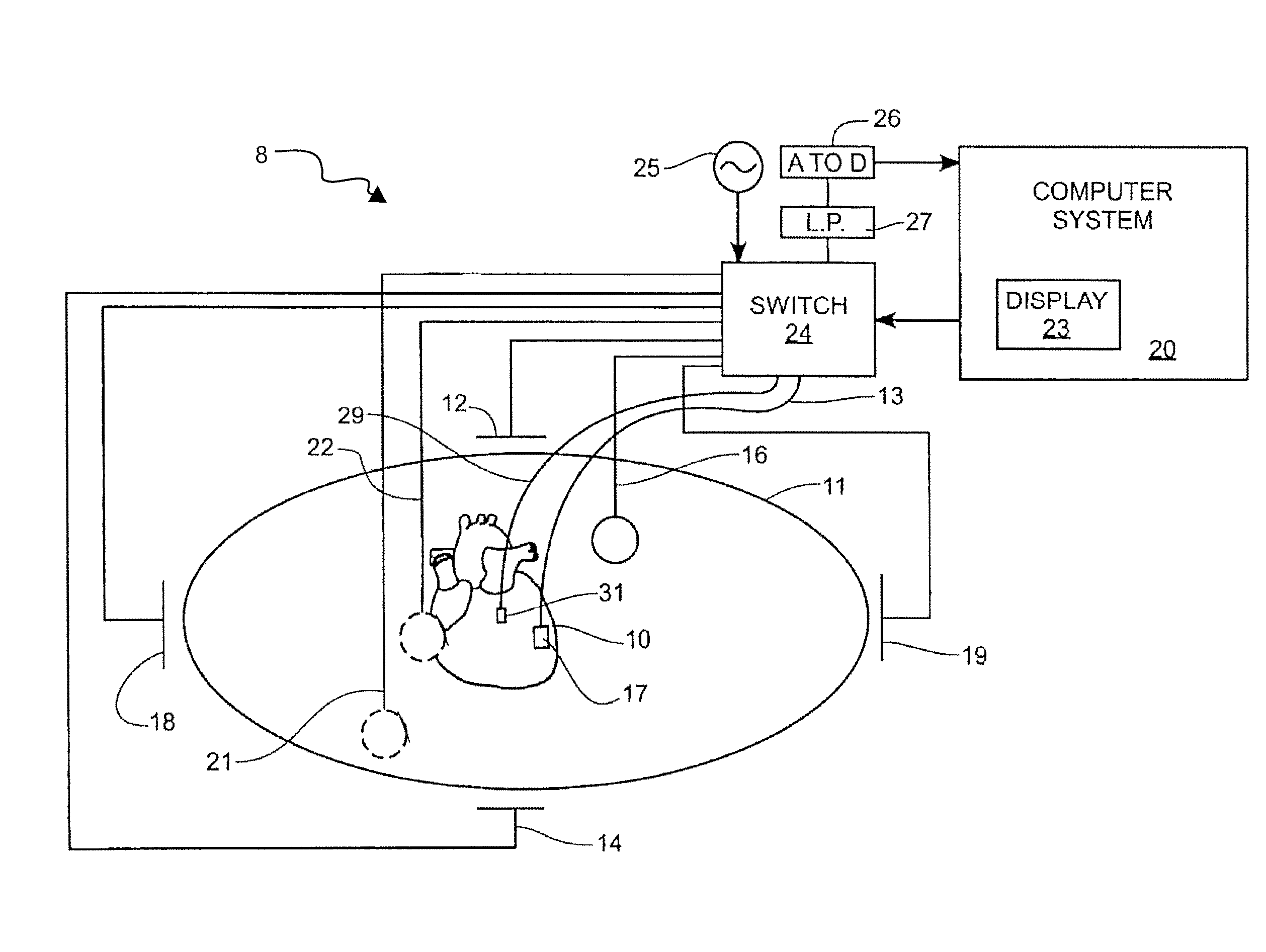
[0026]FIG. 1 shows a schematic diagram of a system 8 according to the present invention for conducting cardiac electrophysiology studies by measuring electrical activity occurring in a heart 10 of a patient 11 and three-dimensionally mapping the electrical activity and / or information related to the electrical activity. In one embodiment, for example, the system 8 can instantaneously locate up to sixty-four electrodes in and / or around a heart and the vasculature of a patient, measure electrical activity at up to sixty-two of those sixty-four electrodes, and provide a three-dimensional map of time domain and / or frequency domain information from the measured electrical activity (e.g., electrograms) for a single beat of the heart 10. The number of electrodes capable of being simultaneously monitored is limited only by the number of electrode lead inputs into the system 8 and the processing speed of the system 8. The electrodes ma...


PUM


Abstract

An electrophysiology apparatus is used to measure electrical activity occurring in a heart of a patient and to visualize the electrical activity and / or information related to the electrical activity. A three-dimensional map of the electrical activity and / or the information related to the electrical activity is created. Exemplary maps include a time difference between action potentials at a roving electrode and a reference electrode, the peak-to-peak timing of action potentials at the roving electrode, the peak negative voltage of action potentials at the roving electrode, complex fractionated electrogram information, a dominant frequency of an electrogram signal, a maximum peak amplitude at the dominant frequency, a ratio of energy in one band of the frequency-domain to the energy in a second band of the frequency-domain, a low-frequency or high-frequency passband of interest, a frequency with the maximum energy in a passband, a number of peaks within a passband, an energy, power, and / or area in each peak, a ratio of energy and / or area in each peak to that in another passband, and a width of each peak in a spectrum. Colors, shades of colors, and / or grayscales are assigned to values of the parameters and colors corresponding to the parameters for the electrograms sampled by the electrodes are updated on the three-dimensional model.

Description

BACKGROUND OF THE INVENTION[0001]1. Field of the Invention[0002]The instant invention relates to an electrophysiology apparatus used to measure electrical activity occurring in a heart of a patient and to visualize the electrical activity and / or information related to the electrical activity. In particular, the instant invention relates to three-dimensional mapping of the electrical activity and / or the information related to the electrical activity.[0003]2. Background Art[0004]The heart contains two specialized types of cardiac muscle cells. The majority, around ninety-nine percent, of the cardiac muscle cells is contractile cells, which are responsible for the mechanical work of pumping the heart. Autorhythmic cells comprise the second type of cardiac muscle cells, which function as part of the autonomic nervous system to initiate and conduct action potentials responsible for the contraction of the contractile cells. The cardiac muscle displays a pacemaker activity, in which membra...

Claims


Application Information

Patent Timeline
Patent Type & Authority Patents(United States)
IPC IPC(8): A61B5/02A61B5/05
CPCA61B5/04012A61B5/0452A61B5/044A61B5/0422A61B5/316A61B5/339A61B5/287A61B5/349A61B5/367
Inventor AFONSO, VALTINO X.BELHE, KEDAR RAVINDRASCHWEITZER, JEFFREY A.
Owner ST JUDE MEDICAL ATRIAL FIBRILLATION DIV
Learn More