Systems and methods for evaluating ventricular dyssynchrony using atrial and ventricular pressure measurements obtained by an implantable medical device

Active Publication Date: 2011-09-13
PACESETTER INC
View PDF56 Cites 21 Cited by
  • Summary
  • Abstract
  • Description
  • Claims
  • Application Information

AI Technical Summary

Benefits of technology

[0020]The interventricular mechanical delay is the primary parameter representative of ventricular dyssynchrony. In one example, to determine the interventricular mechanical delay and thereby evaluate ventricular dyssynchrony, the device detects LAP using a pressure sensor implanted in or on the left atrium and then identifies the c-wave therein. The device also detects the RVP using a pressure sensor implanted in or on the RV and identifies an upstroke point therein. The interventricular mechanical delay is then determined based on the time interval between the upstroke of the RVP and the c-wave of the LAP. By determining the interventricular delay based only on LAP and RVP, no LVP sensor is required. Alternatively, if an LVP sensor is also implanted, the LAP-based technique provides an independent procedure for assessing the interventricular delay (in addition to any LVP / RVP measurements) so as to provide a more robust overall determination of the interventricular delay.
[0024]Hence, the various ventricular mechanical and electromechanical parameters are conveniently detected by an implantable medical device based LAP measurements, alone or in combination with other signals, such as LV and RV IEGM signals as well as RVP measurements. None of the parameters requires detection of LVP by the implanted device. These measurement techniques may be advantageously employed alone or in combination with other measurement techniques to provide for a robust overall ventricular dyssynchrony evaluation procedure. Preferably, therapy delivered by the implanted device is controlled based on the various detected parameters. For example, the interventricular pacing delay (i.e. the VL-pulse-VR pulse pacing delay) may be automatically adjusted in accordance with CRT techniques so as to reduce the interventricular mechanical delay. If the patient is know to have diastolic heart failure, pacing may be delivered so as to reduce the measured electromechanical delays while also reducing the LV and RV systolic intervals. Appropriate diagnostic information pertaining to the various detected parameters is preferably stored within the implanted device for subsequent review by the clinician or relayed directly to the clinician via a bedside monitor network or other suitable communication network. The diagnostic information may also be used to track the progression or regression of heart failure or other cardiovascular diseases.

Problems solved by technology

Typically, the heart loses propulsive power because the cardiac muscle loses capacity to stretch and contract.
Often, the ventricles do not adequately eject or fill with blood between heartbeats and the valves regulating blood flow become leaky, allowing regurgitation or back-flow of blood.
The impairment of arterial circulation deprives vital organs of oxygen and nutrients.
Fatigue, weakness and the inability to carry out daily tasks may result.
As heart failure progresses, it tends to become increasingly difficult to manage.
Even the compensatory responses it triggers in the body may themselves eventually complicate the clinical prognosis.
If the oxygen supply falls short of the growing demand, as it often does, further injury to the heart may result.
The additional muscle mass may also stiffen the heart walls to hamper rather than assist in providing cardiac output.
A particularly severe form of heart failure is congestive heart failure (CHF) wherein the weak pumping of the heart leads to build-up of fluids in the lungs and other organs and tissues.
Heart failure is often associated with electrical signal conduction defects within the heart.
Any of these conduction pathways may potentially be degraded.
The electrical impulse continues to be conducted to the postero-lateral wall of the left ventricle causing its activation and depolarization but, due to an inability to use the native conduction system, this activation and contraction is delayed.
LBBB thus results in an abnormal activation of the left ventricle inducing desynchronized ventricular contraction (i.e. ventricular dyssynchrony) and impairment in cardiac performance.
However, this evaluation and programming requires an office visit and is therefore a timely and expensive process.
It also restricts the evaluation to a resting state, commonly with the patient in a supine position.
As such, the system is not necessarily optimized for activity, for the upright position, for other times of day since there may also be a circadian rhythm to cardiac function.
Moreover, when relying on any external hemodynamic monitoring system, the control parameters of the pacemaker or ICD cannot be automatically adjusted to respond to on-going changes in patient cardiac function.
Unfortunately, conventional pacemakers and ICDs are not capable of detecting the aforementioned mechanical and electromechanical parameters.
That is, although conventional devices are certainly capable of sensing electrical cardiac signals such as P-waves, R-waves, and T-waves, the devices are not typically capable of sensing actual mechanical contraction of the various chambers of the heart and hence cannot detect the mechanical and electromechanical parameters needed for optimal CRT control.
Although the techniques of Min et al. are quite useful, the various parameters specific to ventricular dyssynchrony noted above are not detected by those techniques.
However, reliable detection of LVP is problematic and so it would be desirable to provide alternative techniques for determining the interventricular delay that do not necessarily require an LVP sensor.
Also, Struble et al. does not appear to provide techniques for detecting each of the various other ventricular mechanical and electromechanical parameters listed above by exploiting pressure measurements.
However, Mann et al. does not appear to set forth techniques for measuring the interventricular delay as well as the various other ventricular mechanical and electromechanical intervals listed above that are pertinent to ventricular dyssynchrony.

Method used


Image

  • Systems and methods for evaluating ventricular dyssynchrony using atrial and ventricular pressure measurements obtained by an implantable medical device
  • Systems and methods for evaluating ventricular dyssynchrony using atrial and ventricular pressure measurements obtained by an implantable medical device
  • Systems and methods for evaluating ventricular dyssynchrony using atrial and ventricular pressure measurements obtained by an implantable medical device

Examples


Embodiment Construction

[0040]The following description includes the best mode presently contemplated for practicing the invention. This description is not to be taken in a limiting sense but is made merely to describe general principles of the invention. The scope of the invention should be ascertained with reference to the issued claims. In the description of the invention that follows, like numerals or reference designators are used to refer to like parts or elements throughout.

Overview of Implantable Medical System

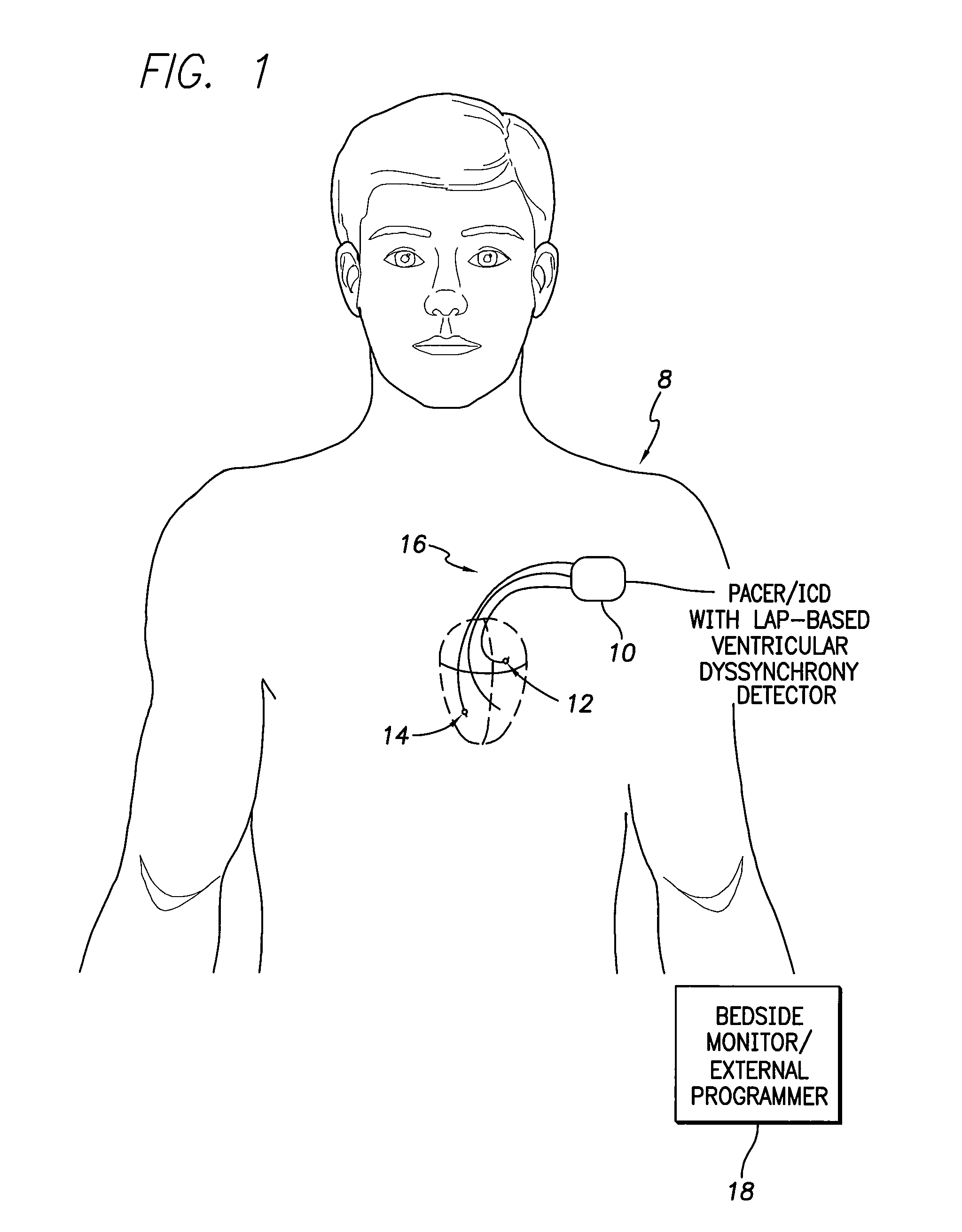
[0041]FIG. 1 illustrates an implantable medical system 8 capable of detecting parameters representative of ventricular dyssynchrony based, in part, on LAP measurements and also for controlling delivery of appropriate therapy in response thereto. To this end, a pacer / ICD 10 (or other implantable medical device) receives signals representative of LAP from an LAP sensor 12 implanted in or on the left atrium or from any other implanted device capable of detecting LAP (such as an epicardial LAP de...


PUM


Abstract

Techniques are provided for exploiting left atrial pressure (LAP) measurements to determine various ventricular mechanical and electromechanical contraction intervals and for evaluating ventricular dyssynchrony based on those intervals. In one example, LAP measurements are combined with right ventricular pressure (RVP) measurements to determine the interventricular mechanical delay, which is the primary parameter representative of ventricular dyssynchrony. Other intervals determined based, in part, on LAP measurements include the electromechanical delay for the left ventricle (LV), as well as the LV systolic and diastolic intervals. Techniques are also described herein for detecting various RV intervals including the electromechanical delay for the RV, as well as the RV systolic and diastolic intervals.

Description

FIELD OF THE INVENTION[0001]The invention generally relates to implantable medical devices, such as pacemakers or implantable cardioverter / defibrillators (ICDs), and in particular to techniques for evaluating ventricular dyssynchrony within heart failure patients and for controlling therapy in response thereto.BACKGROUND OF THE INVENTION[0002]Heart failure is a debilitating disease in which abnormal function of the heart leads in the direction of inadequate blood flow to fulfill the needs of the tissues and organs of the body. Typically, the heart loses propulsive power because the cardiac muscle loses capacity to stretch and contract. Often, the ventricles do not adequately eject or fill with blood between heartbeats and the valves regulating blood flow become leaky, allowing regurgitation or back-flow of blood. The impairment of arterial circulation deprives vital organs of oxygen and nutrients. Fatigue, weakness and the inability to carry out daily tasks may result. Not all heart...

Claims


Application Information

Patent Timeline
IPC IPC(8): A61B5/02
CPCA61B5/02158A61B5/349A61B5/021A61B5/02108A61M2230/04
Inventor SHELCHUK, ANNE M.
Owner PACESETTER INC
Why Eureka
Try Eureka