
Viable tissue repair implants and methods of use

a tissue repair and implant technology, applied in the field of tissue repair and implant augmentation, can solve the problems of inconvenient use, inconvenient use, and high labor intensity, and achieve the effects of reducing manipulation time, repairing tissue injury or defect at minimal cost, and without cell isolation and amplification

Inactive Publication Date: 2011-03-08
DEPUY SYNTHES PROD INC
View PDF312 Cites 91 Cited by
  • Summary
  • Abstract
  • Description
  • Claims
  • Application Information

AI Technical Summary

Benefits of technology

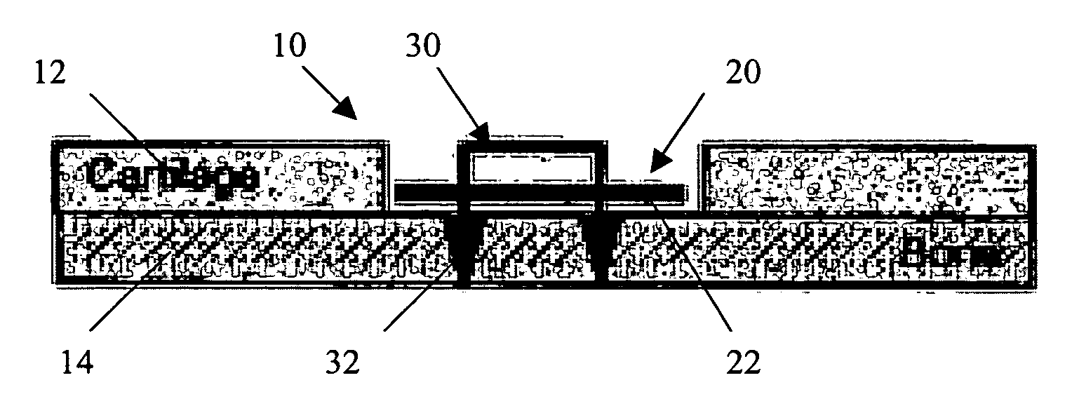
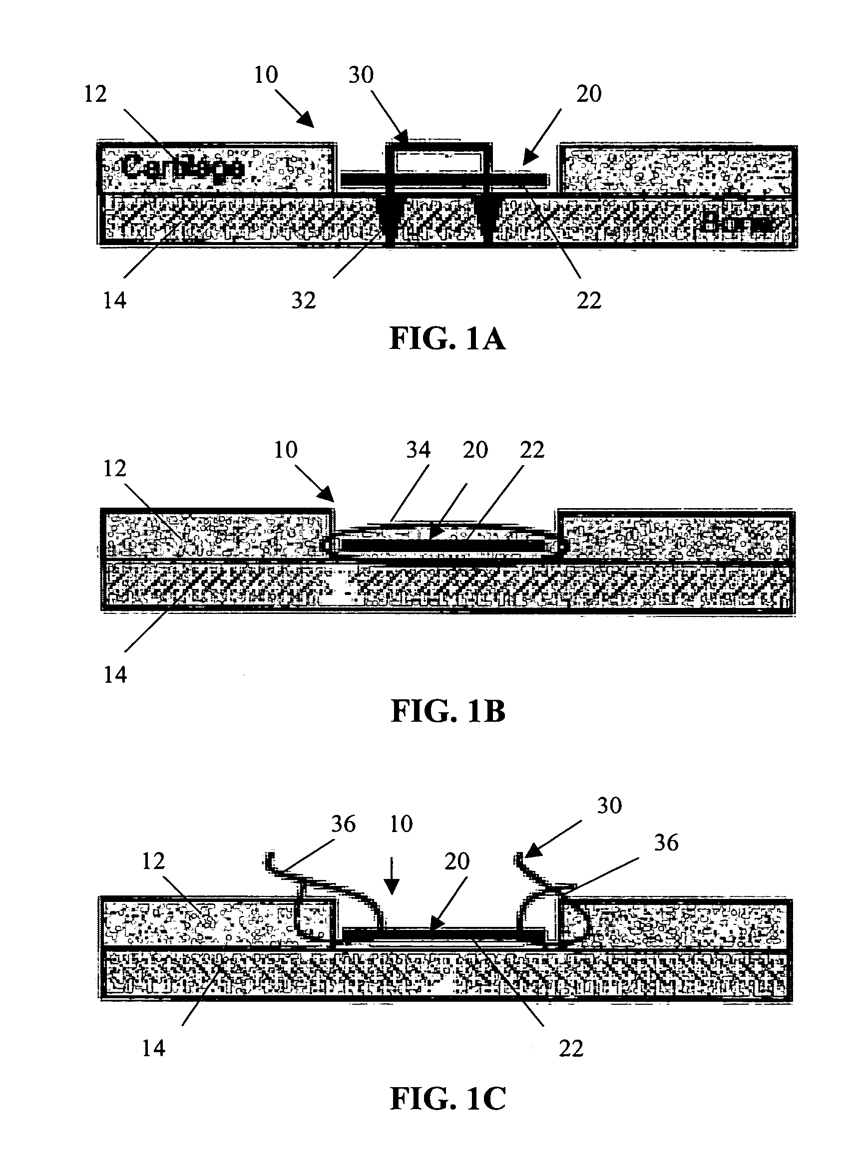
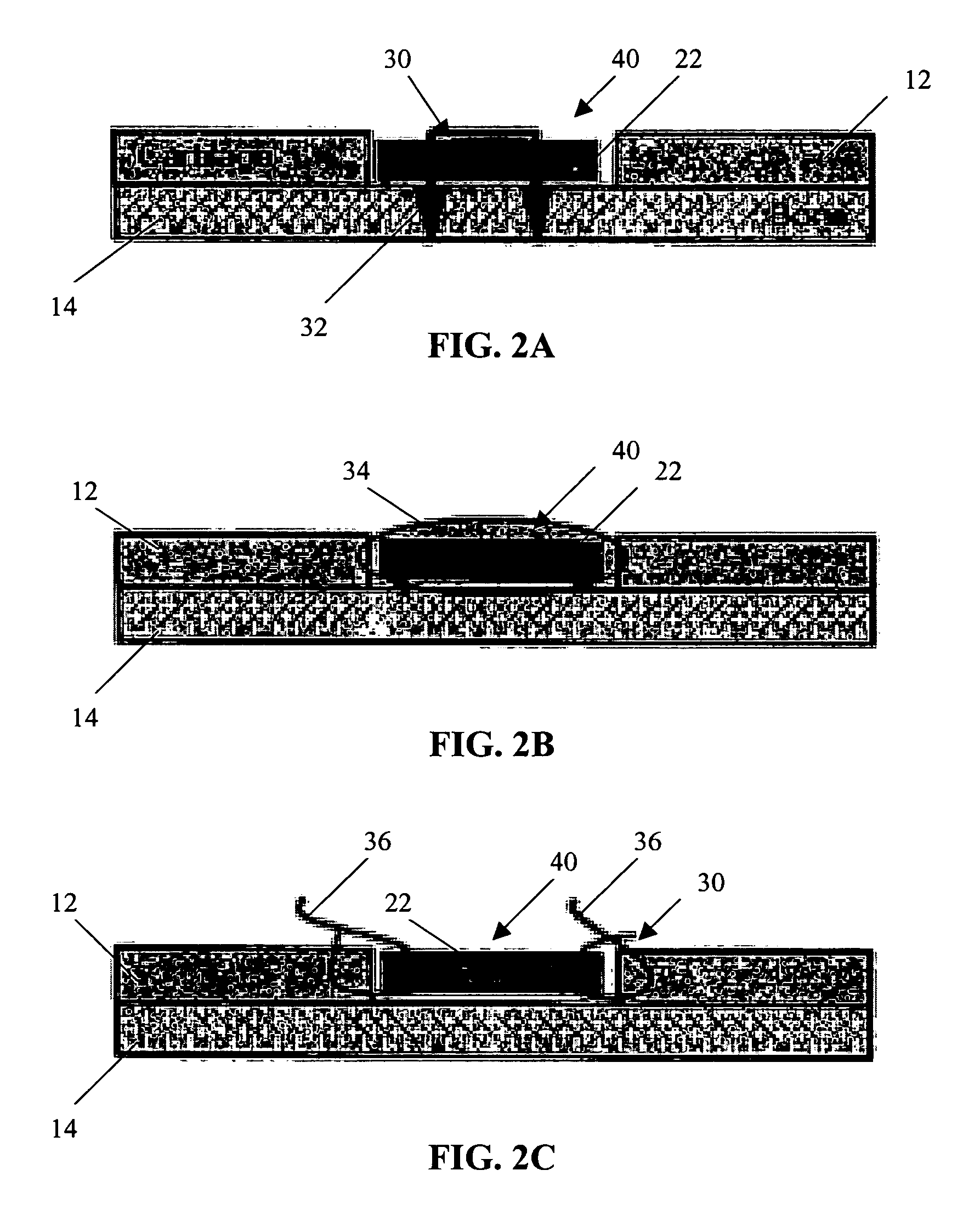
The present invention provides a biocompatible tissue implant for repairing tissue defects or injuries. The implant is made from a biological tissue slice that contains viable cells capable of tissue regeneration and repair. The tissue slice can be harvested from healthy tissue during the surgery and is suitable for implantation at the injury or defect site. The implant can be secured to the site with a retaining element and can be used for tissue repair in various orthopaedic surgeries. The use of a thin tissue slice provides a native tissue surface with similar mechanical properties to that of neighboring tissue and allows for better retention of the cells at the injury or defect site. The cells have the ability to migrate out from the tissue and provide good integration between the implanted tissue and the injury or defect site. The invention provides a cost-effective and efficient method for repairing tissue defects or injuries with minimal manipulation time and improved cell viability.

Problems solved by technology

While the trend towards using tissue engineering approaches to tissue repair continues to gain popularity, mainly because of the long-term benefits provided to the patient, these current techniques are not without drawbacks.
One disadvantage with current tissue engineering techniques is that they can be time consuming.
This manual, labor-intensive process is extremely costly and time consuming.
Although the clinical data suggest long-term benefits for the patient, the prohibitive cost of the procedure, combined with the traumatic impact of two surgical procedures, has hampered adoption of these techniques.
This model is limited in application because of the secondary surgery and high costs involved.
And though allografts have been used for tissue repair in the past, this solution is also not ideal because of the limited availability of graft material and the potential for disease transmission.

Method used


Image

  • Viable tissue repair implants and methods of use
  • Viable tissue repair implants and methods of use
  • Viable tissue repair implants and methods of use

Examples


example 1

[0116]In this in vitro study, cellular migration and new matrix formation from minced and shredded bovine anterior cruciate ligament (ACL) tissue into non-woven tissue scaffold (PANACRYL) was evaluated and compared. Pre-scored and sterilized PANACRYL non-woven sheets were trimmed to yield two (2) 2.5×2 cm sheets. Next, bovine ACL tissue samples were obtained from two knees from the same animal. To prepare the shredded tissue, an isolated section of the bovine ACL was trimmed under aseptic conditions to measure approximately 2×2×0.5 cm in overall dimensions. Using a sterile scalpel, multiple full thick incisions were made parallel to the fibers of the ACL section, yielding tissue strands measuring approximately 2 cm in length and 0.1 cm in maximum diameter. The tissue strands were placed parallel to the long axis of a PANACRYL sheet to form a composite implant. To prepare the control, minced ACL tissue fragments were also applied to a sheet of PANACRYL. The minced tissue fragments we...

example 2

[0127]In this in vitro study, sliced meniscal tissue was tested as a source of viable cells for meniscal regeneration. First, an isolated bovine meniscus was obtained and trimmed to remove the surrounding synovium. Using a sterile dermatome, slices of meniscus were removed. The thickness of the slices were either 200 μm, 300 μm or 500 μm. The slices were approximately 1 to 2 cm in length. The meniscal slices were seeded onto scaffolds comprising sterilized, 65:35 polyglycolic acid / poly caprolactone acide foam reinforced with polydioxone mesh at a density of 20 mg / cm2. The scaffolds measured 4×2.5 cm. Platelet rich plasma (PRP) was added to the scaffolds at a concentration of 20 μl / cm2 and the scaffolds cultured for 3 and 5 weeks in Dulbecco's modified eagles medium (DMEM) supplemented with 0.5% fetal bovine serum (FBS). After 3 and 5 weeks, the samples were prepared for histological evaluation. Sections of the samples were obtained and stained with hematoxylin and eosin.

[0128]Result...

example 3

[0132]In this in vitro study, minced tissue fragments were used in conjunction with mosaicplasty techniques to demonstrate that better integration between cartilage plugs can be achieved and cartilage repair of damaged tissue can be enhanced by the addition of minced cartilage fragments.

[0133]Healthy articular cartilage was obtained from bovine stifle. A 3 mm biopsy punch was used to punch cylinders or plugs of cartilage tissue. The rest of the cartilage tissue, which was substantially free of bone, was minced using scalpel blades to obtain small tissue fragments. The size of the tissue fragments varied but was less than or equal to 1×1 mm in dimension. Four 3 mm cartilage cylinders were placed together in parallel to each other longitudinally in a glass cylinder with an inner diameter of 8 mm. In one group, a blood clot was then formed inside the glass cylinder to keep the tissues together. In another group, the minced cartilage tissue was placed in the glass cylinder with four 3 m...


PUM

PropertyMeasurementUnit
thicknessaaaaaaaaaa
thicknessaaaaaaaaaa
thicknessaaaaaaaaaa

Abstract

Biocompatible tissue implants are provided for repairing a tissue injury or defect. The tissue implants comprise a biological tissue slice that serves as a source of viable cells capable of tissue regeneration and / or repair. The biological tissue slice can be harvested from healthy tissue to have a geometry that is suitable for implantation at the site of the injury or defect. The harvested tissue slice is dimensioned to allow the viable cells contained within the tissue slice to migrate out and proliferate and integrate with tissue surrounding the injury or defect site. Methods for repairing a tissue injury or defect using the tissue implants are also provided.

Description

CROSS REFERENCE TO RELATED APPLICATIONS[0001]Not applicable.STATEMENT REGARDING FEDERALLY SPONSORED RESEARCH[0002]Not applicable.FIELD OF THE INVENTION[0003]The present invention relates to methods and apparatus for the treatment of tissue injuries or defects. Specifically, the present invention relates to tissue repair and augmentation implants, and more particularly, to tissue implants having viable cells capable of tissue regeneration and integration with tissue surrounding the area to be repaired, as well as methods for using such tissue implants.BACKGROUND OF THE INVENTION[0004]Injuries to tissue, such as cartilage, skin, muscle, bone, tendon, and ligament where the tissue has been injured or traumatized frequently require surgical intervention to repair the damage and facilitate healing. Such surgical repairs can include suturing or otherwise repairing the damaged tissue with known medical devices, augmenting the damaged tissue with other tissue, using an implant, a graft or a...

Claims


Application Information

Patent Timeline
Patent Type & Authority Patents(United States)
IPC IPC(8): A61F2/02A61F2/08A61F2/28A61L27/00A61L27/38
CPCA61L27/3683A61L27/38A61L2430/06A61P17/02A61P19/02
Inventor HARMON, ALEXANDER M.KLADAKIS, STEPHANIE M.HWANG, JULIA
Owner DEPUY SYNTHES PROD INC
Learn More