
Active suture for the delivery of therapeutic fluids

a technology of active sutures and therapeutic fluids, applied in the field of functional sutures, can solve the problems of not being safe to deliver, not demonstrating the form and function of a device that could cost-effectively facilitate localized delivery of therapeutic fluids to the wound site over an extended period of time, and not providing efficacy

Active Publication Date: 2011-01-25
ETHICON INC
View PDF31 Cites 792 Cited by
  • Summary
  • Abstract
  • Description
  • Claims
  • Application Information

AI Technical Summary

Benefits of technology

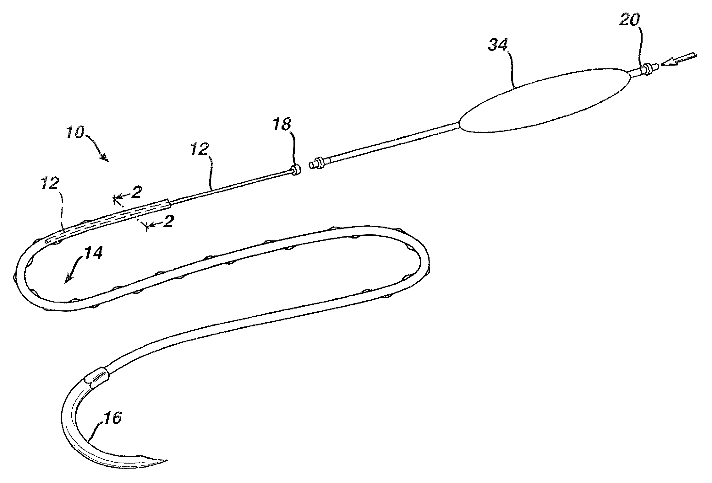
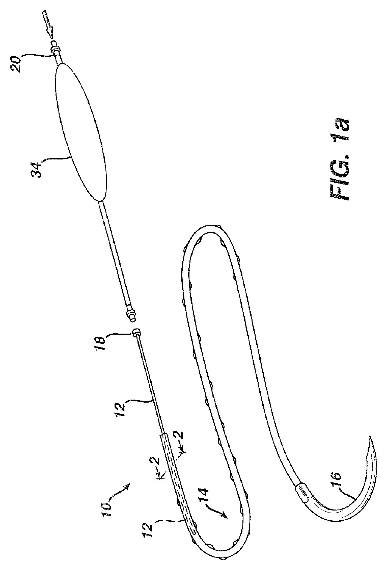
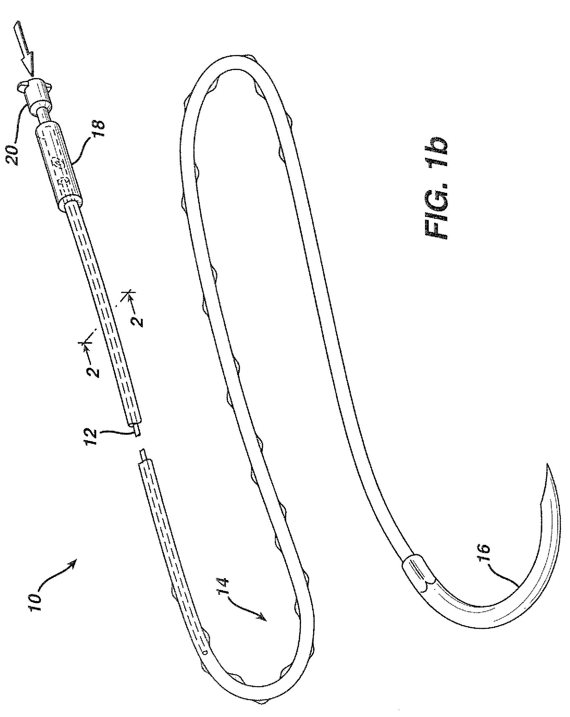
The patent describes an active suture that can be used to close wounds and administer fluids to them. The suture has a braided structure with a passageway embedded within it. The passageway has a smaller diameter than the braided suture and is connected to the braided suture at one end. The other end of the passageway has an opening. The method of use involves attaching a surgical needle to the end of the braided suture and connecting a connector to the other end of the passageway. This allows for the delivery of fluids to the wound without compromising the suture's ability to close it. The technical effect of this invention is the creation of a versatile suture that can be used for both wound closure and fluid delivery.

Problems solved by technology

However, the form and function of a device that could cost-effectively facilitate localized delivery of therapeutic fluids directly to the wound site over an extended period of time are not apparent.
However, in many cases, the increased concentration of medication that may provide the most efficacious result at the wound site, may not be safely delivered through an IV since toxic side effects may occur at various organs within the body.
Other medications, such as certain local anesthetics, only provide an efficacious result when delivered locally and are simply not compatible with IV delivery methods.
Although the syringe and hypodermic needle provide a means for localized drug delivery, the continuous delivery of medication via injection over an extended time period is not practical.
Indeed, over time the medication dissipates to a concentration below that required to achieve a therapeutic effect and additional injections must be prescribed.
The patient may suffer discomfort and repetitive disturbance if multiple injections must be repeatedly administered.
However, a greater risk of infection and compromised wound healing may be associated with this deployment method since the infusion catheter may serve as a pathway for pathogens to enter the surgical incision.
Although these catheters provide a means of continuously delivering a therapeutic fluid to the wound, a number of drawbacks exists.
Even so, the infusion catheter may not be firmly anchored and accidental removal of the catheter from the wound site by the patient is not uncommon.
Finally, there are significant additional costs, ranging from hundreds to thousands of dollars, associated with the use of these specialized catheters and the supporting reservoirs and pumps that must be employed for their operation.
Further, since the suture must be present in most cases to achieve wound closure, the number of invasive procedures that a patient must suffer is not necessarily increased.
Moreover the flexibility exhibited by a suture is considerable greater than the flexibility exhibited by infusion catheters, consequently, the suture may be placed in a complicated pattern or in locations that would be hard to reach with a conventional infusion catheter.
First of all, monofilament sutures are flaw sensitive.
The introduction of pores or perforations into the wall of the hollow suture may result in a substantial decrease in the strength performance of the suture and lead to its inability to insure secure closure of the wound.
Secondly, the amount of medicine that may be contained inside of a hollow suture is small.
Active control of the drug delivery rate and continuous drug delivery are not possible.

Method used


Image

  • Active suture for the delivery of therapeutic fluids
  • Active suture for the delivery of therapeutic fluids
  • Active suture for the delivery of therapeutic fluids

Examples


example 1

[0044]In order to demonstrate the ability of the active suture to distribute a fluid to surrounding tissue, a PET braided suture, containing a polypropylene tube that terminates within the braided suture, as depicted in FIGS. 1b, and 4a, was employed in an in vitro experiment wherein the active suture was passed multiple times though gelatin and subsequently connected to an IV delivery system that delivered water containing a blue pigment to the portion of the active suture that was imbedded in the gelatin. A series of time-elapsed images are shown in FIGS. 10a, 10b and 10c. FIG. 10a, taken at the onset of the experiment, shows the active suture 70 embedded in gelatin 72. The black mark on the active suture 74 indicates the location at which the internal passageway terminates. As time progresses, the pigment 76 spreads out around the active suture as shown in FIG. 10b. Ultimately, as shown in FIG. 10c, the fluid spreads to encompass the entire region surrounding the wound.

example 2

[0045]The incorporation of internal passageways into the active sutures should not compromise the tensile strength and knot tensile strength of the sutures to below standard acceptable levels if the active suture is to be used for both wound approximation and fluid infusion. The knot tensile strengths of PET braided sutures in United States Pharmacopia (USP) standard sizes of 0 and 2 that have polypropylene tubes imbedded along side their core filaments were measured according to United States Pharmacopia (USP) standard 23. Size 0 sutures contained tubes with outside diameters of approximately 130 μm and inside diameters of ˜75 μm, and size 2 sutures contained tubes with outside diameters of approximately 230 μm and inside diameters of ˜135 μm. For each test, at least 10 samples were tested per USP specifications. The performance of the PET braided sutures containing the polypropylene tubing at their core easily exceeded minimum performance requirements as set by USP standards, with...

example 3

[0046]Experimental data indicates that extruded polymeric tubes produced from polypropylene, with outside diameters ranging from 0.005″ to 0.010″, with Youngs Moduli ranging between 0.1 and 3 GPa, with outside diameters (O.D.s) that are less than 1.7 times that of their inside diameters (I.D.s) will buckle and collapse when the braided sutures in which they are embedded are tied into square knots similar in form to those commonly used in surgical procedures. Similar experiments conducted with polymeric tubes comprised of polyethylene and polytetraflouroethylene tubes with Youngs moduli ranging between 0.1 and 3 GPa with O.D. to I.D. ratios of greater than 2.3 do not collapse completely inside the square knots of the active suture and fluid can indeed be transferred through the knotted portions. For active sutures that will be tied into knots, preferably the ratio of the O.D. to I.D. is greater than 1.7. More preferably, the ratio of the O.D. to I.D. is greater than 2.0. In these exp...


PUM


Abstract

An active suture that can be used for the delivery of therapeutic fluids to the tissue surrounding a wound is disclosed. The active suture may include a connector designed to join a fluid source, such as a syringe, conventional IV delivery system, or infusion pump to an internal passageway that is embedded within a braided suture. The internal passageway may be comprised of a fine polymeric tube and is capable of conducting and emitting a fluid into at least a portion of the braided suture and surrounding tissue. The invention enables delivery of an efficacious volume of drug bearing solution on the order of milliliters per day, provides a high level of fluid delivery rate control enabling the physician to start or stop drug administration at his / her discretion, and offers a means of providing more than one type of medication that may be selected post-surgically in accord with unexpected patient symptoms that may arise.

Description

CROSS-REFERENCE TO RELATED APPLICATION[0001]This application is a continuation-in-part of U.S. application Ser. No. 10 / 727,367 filed Dec. 4, 2003.FIELD OF INVENTION[0002]The present invention relates to devices that may be used to deliver therapeutic fluids to surgical wounds. More particularly the invention relates to functional sutures that may be used to emit therapeutic or bioactive fluids to the tissue surrounding the suture. In particular, the invention relates to a braided suture having an internal passageway capable of conducting a fluid along at least a portion of the length of the suture that may be attached on one end, through a connector, to a fluid source.BACKGROUND OF THE INVENTION[0003]Much benefit could be realized by delivering therapeutic fluids to the direct vicinity of the surgical wound. Reduced pain, enhanced wound healing, and reduced occurrence of surgical site infections are but a few potential benefits. However, the form and function of a device that could ...

Claims


Application Information

Patent Timeline
Patent Type & Authority Patents(United States)
IPC IPC(8): A61B17/04A61B17/06A61B17/20A61M31/00A61M37/00
CPCA61B17/06166A61M37/00A61B17/0469A61B2017/0404A61B2017/0458A61B2017/0464A61B2017/0475A61B2017/06185
Inventor CICHOCKI, JR., FRANK R.
Owner ETHICON INC
Learn More