Fetal methylation markers
a technology of methylation markers and fetal bodies, which is applied in the field of fetal methylation markers, can solve the problems of presenting an appreciable risk to both the mother and the fetus, invasive conventional methods, etc., and achieves the effect of increasing the risk
- Summary
- Abstract
- Description
- Claims
- Application Information
AI Technical Summary
Benefits of technology
Problems solved by technology
Method used
Image
Examples
example 1
Tumor Suppressor Genes that are Hypermethylated in the Fetus Compared with Maternal Blood
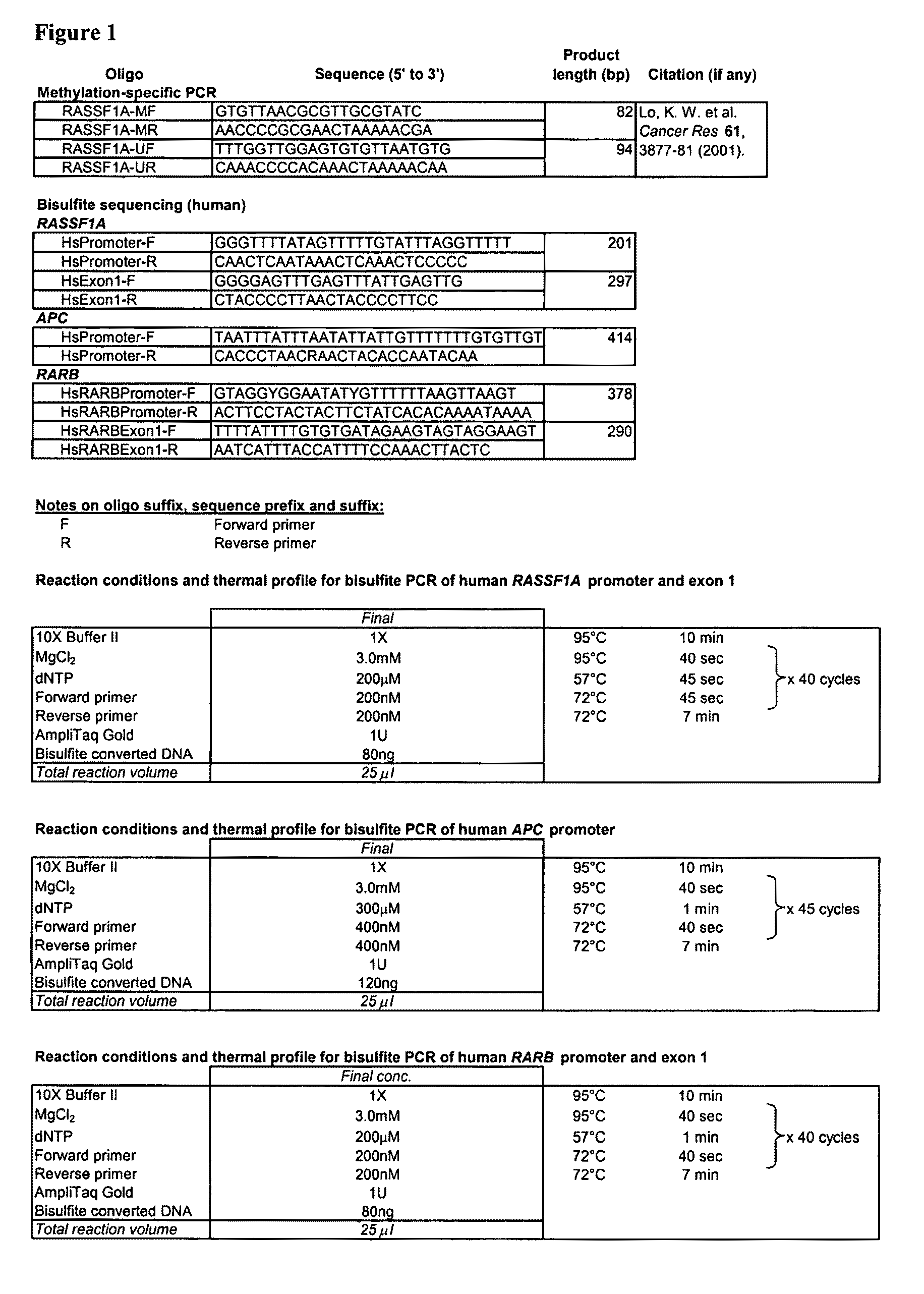
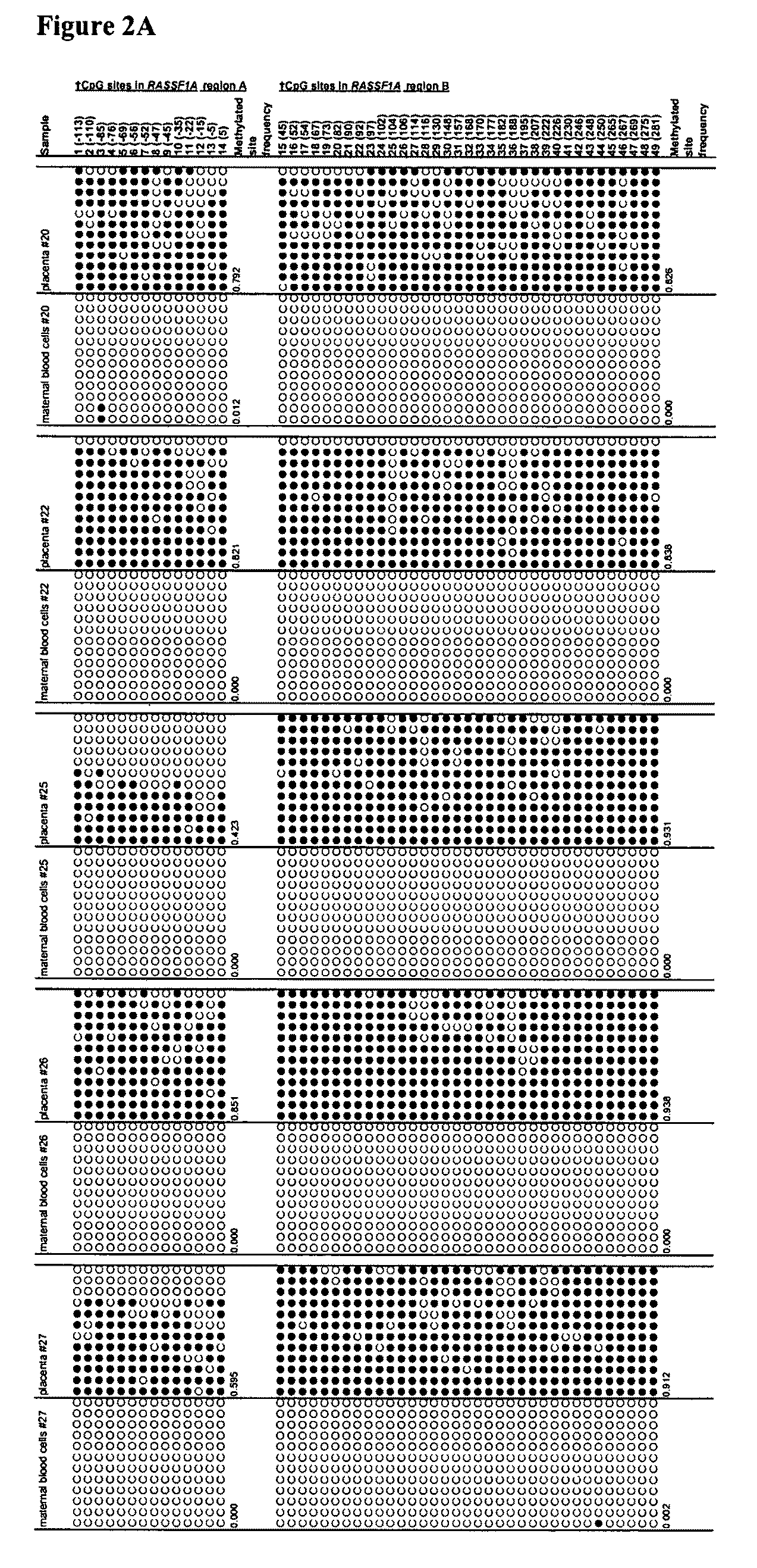
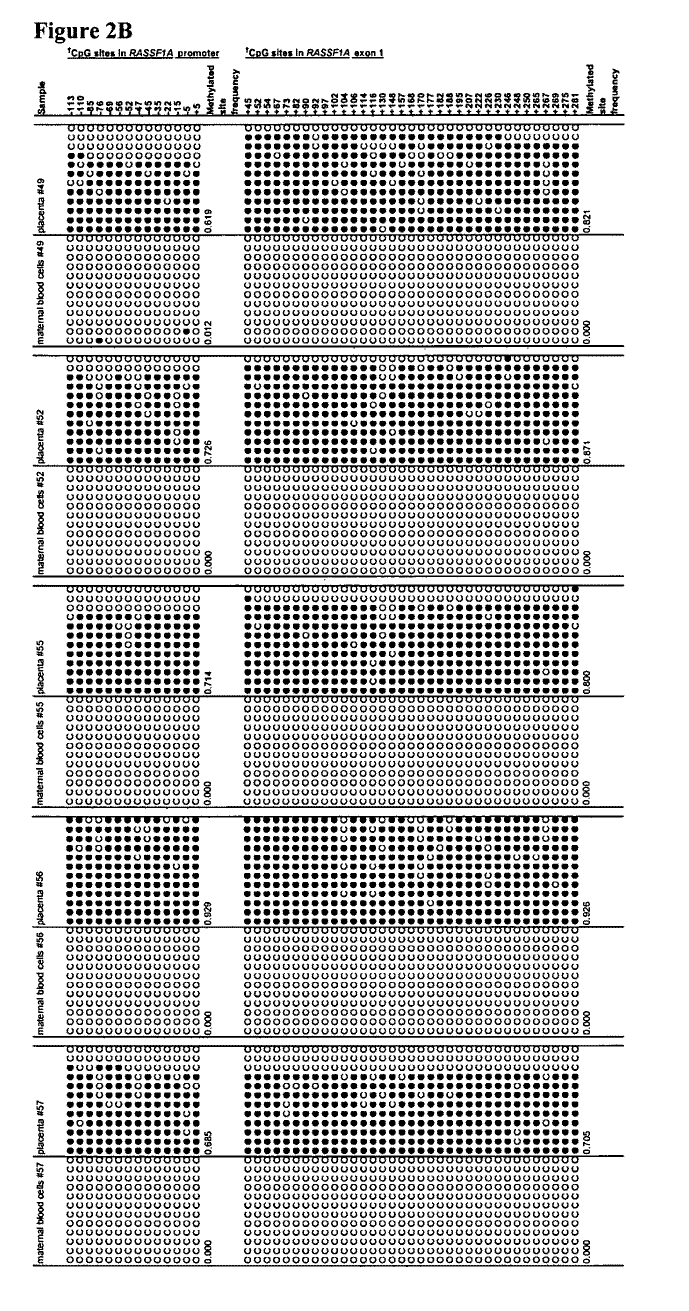
[0080]We aimed to identify epigenetic markers that are fetal-specific in maternal blood. Previous data suggest that fetal DNA molecules in maternal plasma are predominantly derived from the placenta (Chim et al., Proc Natl Acad Sci U S A., 102, 14753-14758; Masuzaki et al., J Med Genet 41, 289-292, 2004; Flori et al., Hum Reprod 19, 723-724, 2004), while the background DNA in maternal plasma may originate from maternal blood cells (Lui et al., Clin Chem 48, 421-427, 2002). Hence, to identify fetal epigenetic markers, the methylation profiles of genomic loci were assessed in both placental tissues and maternal blood cells with an aim to identify loci that demonstrate differential methylation between the two tissue types. Such markers can be used for prenatal diagnosis and monitoring of pregnancy-related conditions.
Subject Recruitment and Sample Collection
[0081]Subjects were recruited from the Dep...
example 2
Methylation Sensitive Enzyme Digestion Followed by Real-Time PCR
[0088]Methylation sensitive enzymes are enzymes that cut DNA only when their recognition site is not methylated. For more description, see, e.g., U.S. patent application publication No. US 2005 / 0158739 (Jeddeloh et al.). For example, BstU I recognizes the CGCG site and cuts the DNA when the CpG is not methylated. As shown in FIG. 8, more than one methylation sensitive enzymes can be used to digest unmethylated DNA, leaving only the methylated sequences intact. Methods such as real-time PCR can be used subsequently to detect the DNA sequences which are still amplifiable after enzymatic digestion.
Materials and Methods
Subject Recruitment and Sample Collection
[0089]Subjects were recruited from the Department of Obstetrics and Gynecology, Prince of Wales Hospital, Hong Kong. The study and the collection of human clinical samples were approved by the institutional review board. Informed consent was sought from each subject. T...
example 3
Detection of RASSF1A in Maternal Plasma After Enzymatic Digestion
[0093]In example 1, by bisulfite sequencing, we have demonstrated that the RASSF1A gene of maternal blood cells is completely unmethylated while that of the placenta (of fetal origin) is heavily methylated. In example 2, we have demonstrated that the RASSF1A sequences from maternal blood cells were completely digested by a methylation sensitive restriction enzyme, while the RASSF1A sequences from placenta were only partially digested by the same enzyme. Plasma DNA is composed of DNA of maternal origin (largely from maternal blood cells and thus is unmethylated) and DNA of fetal origin (placenta being a main contributor and thus is methylated for the markers described in this patent application). It is thus feasible to use methylation sensitive enzyme digestion of plasma DNA to remove the maternal DNA background and to increase the fractional concentration of the fetal-derived RASSF1A molecules in maternal plasma.
[0094]...
PUM
| Property | Measurement | Unit |
|---|---|---|
| molecular weight | aaaaa | aaaaa |
| molecular weight | aaaaa | aaaaa |
| concentrations | aaaaa | aaaaa |
Abstract
Description
Claims
Application Information
 - R&D
- Intellectual Property
- Life Sciences
- Materials
- Tech Scout
- Unparalleled Data Quality
- Higher Quality Content
- 60% Fewer Hallucinations
Browse by: Latest US Patents, China's latest patents, Technical Efficacy Thesaurus, Application Domain, Technology Topic, Popular Technical Reports.
© 2025 PatSnap. All rights reserved.Legal|Privacy policy|Modern Slavery Act Transparency Statement|Sitemap|About US| Contact US: help@patsnap.com


