
Rotating fine needle for core tissue sampling

a core tissue and fine needle technology, applied in medical science, surgery, vaccination/ovulation diagnostics, etc., can solve the problems of difficult aspiration of a sample from a solid mass, inability to remove the necrosis of the pancreas, and traumatic procedures, etc., to facilitate the collection of a core biopsy.

Active Publication Date: 2010-05-25
GRANIT MEDICAL INNOVATION
View PDF19 Cites 97 Cited by
  • Summary
  • Abstract
  • Description
  • Claims
  • Application Information

AI Technical Summary

Benefits of technology

[0012]A related object of the present invention is to provide a needle assembly that requires only a minimal number of needle passes into the organ tissue.
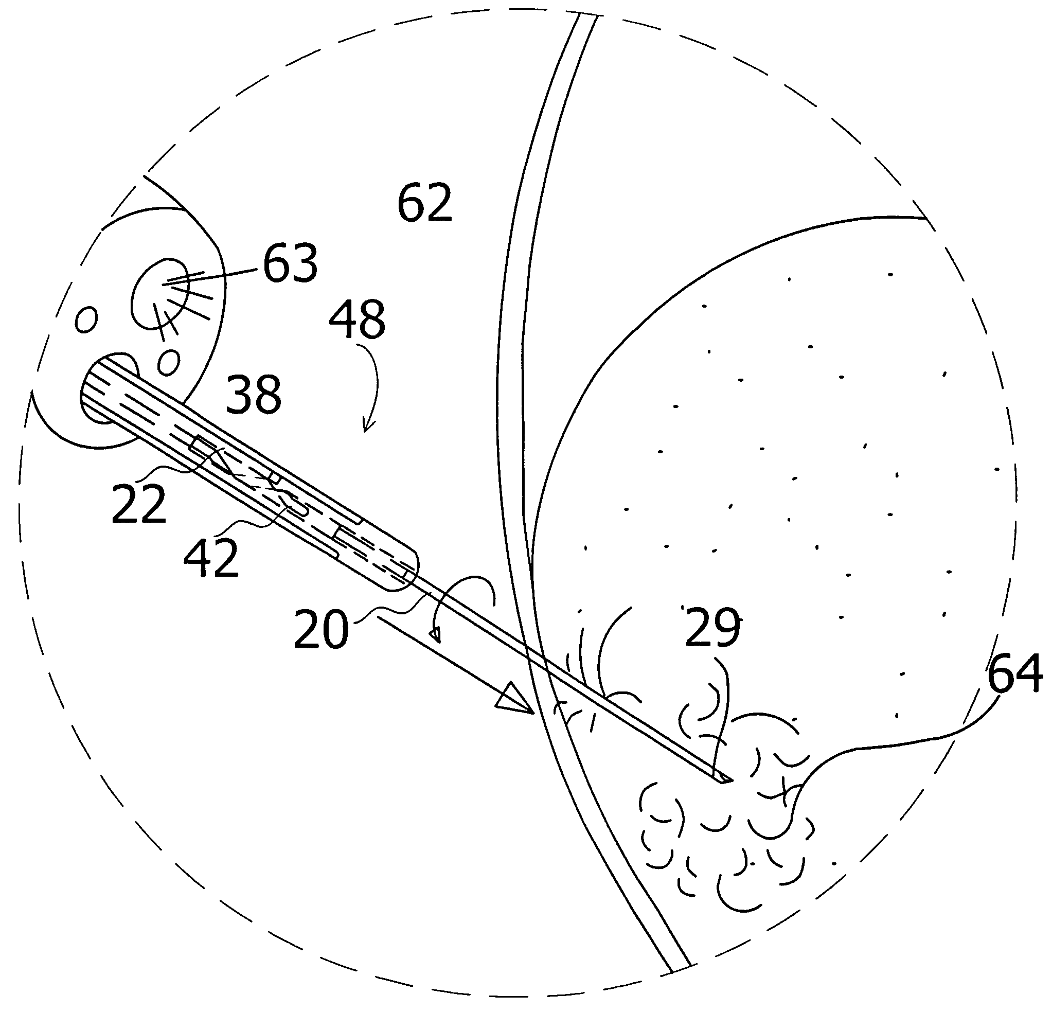
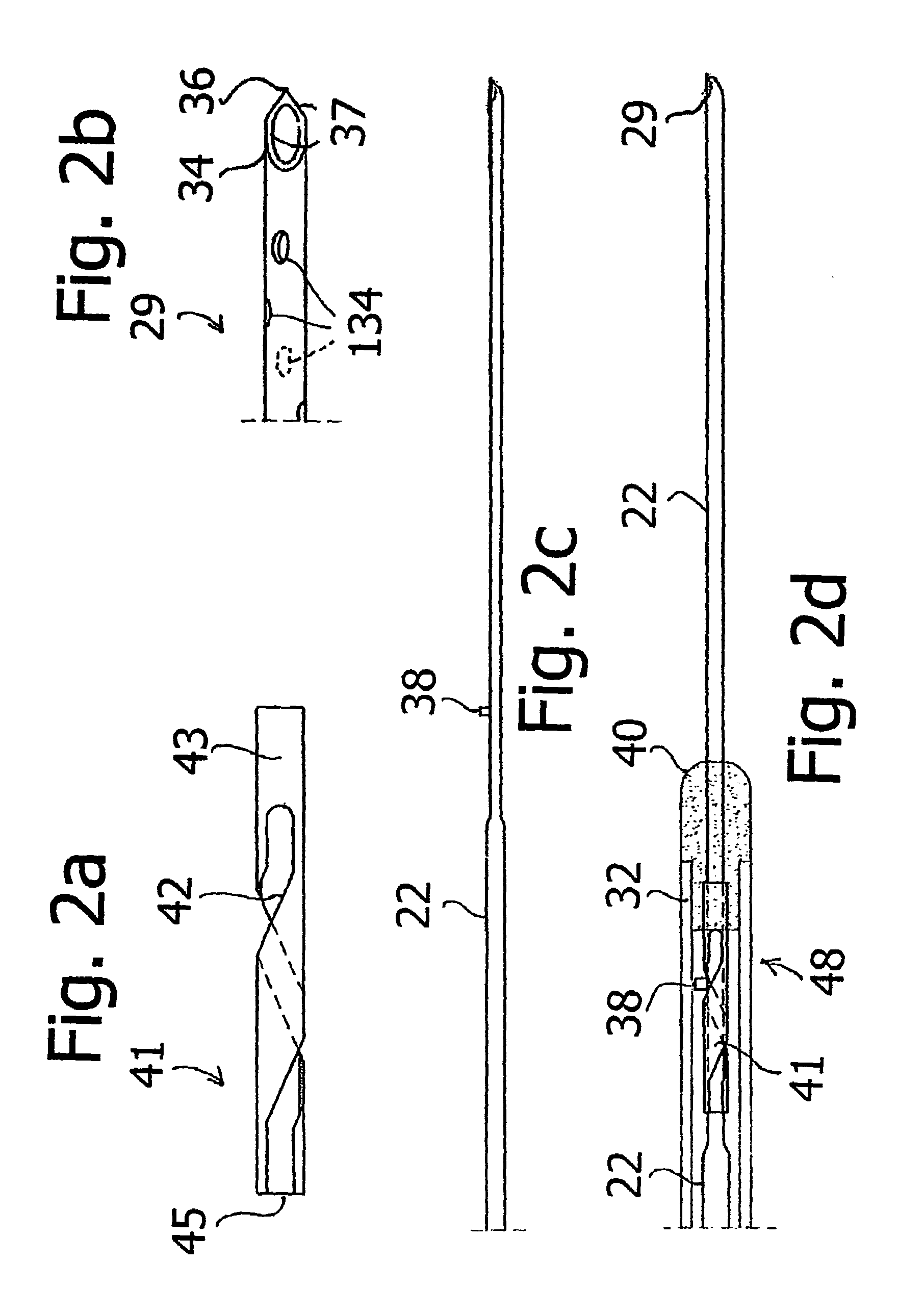
[0028]According to a more specific feature of the present invention, the needle's distal end is configured in the shape of a spoon with a sharp piercing tip and sharp edges extending proximally from the needle tip. Located on the proximal portion of the needle-shaft is a small stub element. In addition, the distal sub-assembly includes a short metal tube, the tube provided with a spiral cutout. The short metal tube is disposed in a fixed state at a distal location of the catheter, affixed to an outer metal collet, the collet providing the most distal portion of the catheter. The needle which is slidably disposed within the catheter is capable of passing freely through the metal tube.

Problems solved by technology

These procedures often result in obtaining a small number of cells with each aspiration, cells which may or may not be diagnostic.
In addition, such procedures are often traumatic because of the multiple needle passes that it necessitates.
When injured, these enzymes are released, and may induce self digestion, and necrosis of the pancreas, and adjacent organs.
Aspirating a sample from a solid mass is difficult.
Even when tumor cells are captured, these are often fragmented, and separated from each other.
It is therefore almost impossible to differentiate a primary pancreatic tumor from a metastatic lesion.
Despite the time consuming and traumatic nature of the current FNA procedure, the consequence of a non-diagnostic aspirate is worse, because a missed diagnosis of pancreatic cancer is a sure death sentence.
While this method is least traumatic with only one needle introduction, it produces a poor yield of diagnostic material.
In the worst case, even these tumor cells are not obtained, only blood cells and normal tissue, necessitating one of the more invasive procedures.
Although these organs are more accessible to the needle than the pancreas, the trauma incurred by a thick core biopsy needle stick is great.
When a woman comes for such a biopsy, she is anxious and afraid of the impending diagnosis.
Oftentimes she leaves the procedure with a large hematoma, an internal blood-clot in a severely bruised breast, resulting from the numerous needle sticks required to obtain cells.
This method has rendered many women even more anxious.

Method used


Image

  • Rotating fine needle for core tissue sampling
  • Rotating fine needle for core tissue sampling
  • Rotating fine needle for core tissue sampling

Examples


Embodiment Construction

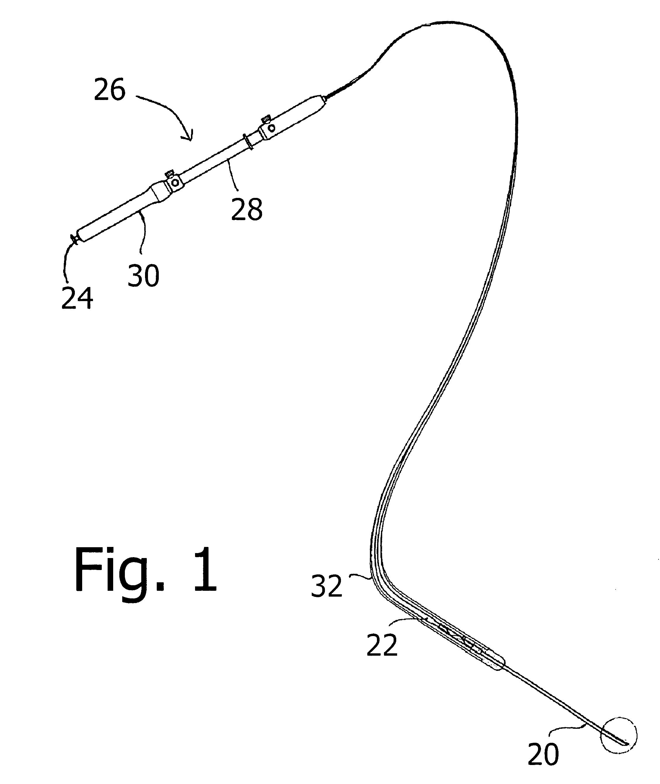
[0051]As illustrated in FIG. 1, a medical instrument assembly for use in procedures for obtaining tissue samples comprises a hollow needle element 20 communicating with a lumen (not shown) of a tubular shaft member 22 to enable suction of a patient's tissue to be obtained by needle 20. Tubular shaft member 22 is provided at a proximal end with a port 24 that communicates with the lumen of the tubular shaft member. As disclosed below with reference to other embodiments, needle element 20 may be an integral distal tip of shaft member 22.

[0052]The instrument assembly of FIG. 1 includes an actuator subassembly 26 including a cylindrical body portion 28 carrying a slidably mounted shifter 30. Shifter is connected to tubular shaft member 22 for moving the tubular shaft member alternatively in the distal direction and the proximal direction through a flexible tubular sheath 32 that is fixed at a proximal end to cylindrical body portion 28. Sheath 32 has a sufficiently small diameter to ena...


PUM


Abstract

A medical instrument usable with an ultrasound-endoscope for performing a needle biopsy on a patient's internal body tissues usable at a surgical site not visible to the unaided eye or viewed endoscopically, comprises an elongate tubular member with a hollow needle element connected at its distal end, a sheath member housing said tubular member and needle element, an actuator subassembly with a shifter member operatively connected to elongate tubular member's proximal end, and a distal camming subassembly, said subassembly enabling a rotating motion of said needle member while handle actuator is moved in the forward direction. Upon inserting an ultrasound-endoscope into a patient and locating a mass, the fine needle with sharply pointed spoon shaped distal end is inserted into the mass aided by endoscopic and ultrasonographic guidance. Once in the mass, a camming action is initiated, causing rotation of the fine needle within the mass, resulting in a scooped out core biopsy. This instrument enables the performance of a fine needle aspiration requiring only one or two needle introductions, with a resultant core biopsy substantial enough for diagnostic purposes.

Description

CROSS-REFERENCE TO RELATED APPLICATION[0001]This application claims the benefit of U.S. Provisional Patent Application No. 60 / 631,348 filed Nov. 29, 2004.FIELD OF THE INVENTION[0002]This invention relates to a medical instrument for use in the application of tissue sampling. An instrument in accordance with this invention is usable with ultrasound, and in some cases an ultrasound-endoscope for performing a needle biopsy operation on a patient's internal body tissues at a surgical site not visible to the unaided eye.BACKGROUND OF THE INVENTION[0003]Fine needle aspiration (FNA) has been a well accepted method for obtaining tissue samples for pathologic or histologic analysis in diagnosing tumors of the pancreas and other soft tissue organs. Endoscopic ultrasound (EUS) and EUS-guided fine needle aspiration (EUS-FNA) have become important tools in the evaluation of pancreatic masses.[0004]Conventional surgical techniques for obtaining tissue samples accessible only through a flexible ul...

Claims


Application Information

Patent Timeline
Patent Type & Authority Patents(United States)
IPC IPC(8): A61B10/00
CPCA61B10/0275A61B17/32053A61B2010/0225A61B17/32002A61B10/0283A61B2017/0046A61B2010/045A61B10/0266A61B10/04
Inventor NAKAO, NAOMI, L.
Owner GRANIT MEDICAL INNOVATION
Learn More