
Device and method for measuring blood flow in an organ

a technology for measuring blood flow and organs, applied in the direction of optical radiation measurement, instruments, catheters, etc., can solve the problems of time-consuming use, difficult implementation, and invasive measurement of input functions, and achieve the effect of less accuracy of evaluation results and greater accuracy in blood flow determination

Active Publication Date: 2009-05-05
UNIV ZURICH +1
View PDF22 Cites 9 Cited by
  • Summary
  • Abstract
  • Description
  • Claims
  • Application Information

AI Technical Summary

Benefits of technology

[0010]It is an object of the present invention to provide a device that can achieve greater accuracy in the determination of the blood flow in an organ, particularly the cerebral blood flow. In particular, the accuracy of the evaluation results should be less dependent on the general conditions, i.e. particularly on how the indicator injection was performed. Furthermore, the device should be non-invasive, and also implementable with a reasonable technical effort / expenditure.

Problems solved by technology

Stroke 6: 245-256), are often difficult to implement, in technical terms, and prove to be time-consuming to use.
Generally, the use of such devices requires the invasive measurement of the input function, i.e. the arterial concentration of the indicator over time.
A significant problem of conventional NIRS devices is that their evaluation units work with a certain inaccuracy, which arises because in the evaluation, until now, the point of departure has been the simplifying assumption that no flow of the indicator out of the system being investigated takes place during the first rise in the input signals (the so-called “rise time”).
Alternative measurement methods, such as positron emission tomography (PET), single-photo photon emission computer tomography (SPECT), or perfusion-weighted magnetic resonance spectroscopy can be implemented technically only by means of extremely expensive devices, and require transport of the patient in question to the measurement unit, in order to be used, and this transport often poses significant risk to the patient; in other words, the devices mentioned cannot be used as so-called “bedside” devices at the patient's bed.

Method used


Image

  • Device and method for measuring blood flow in an organ
  • Device and method for measuring blood flow in an organ
  • Device and method for measuring blood flow in an organ

Examples


Embodiment Construction

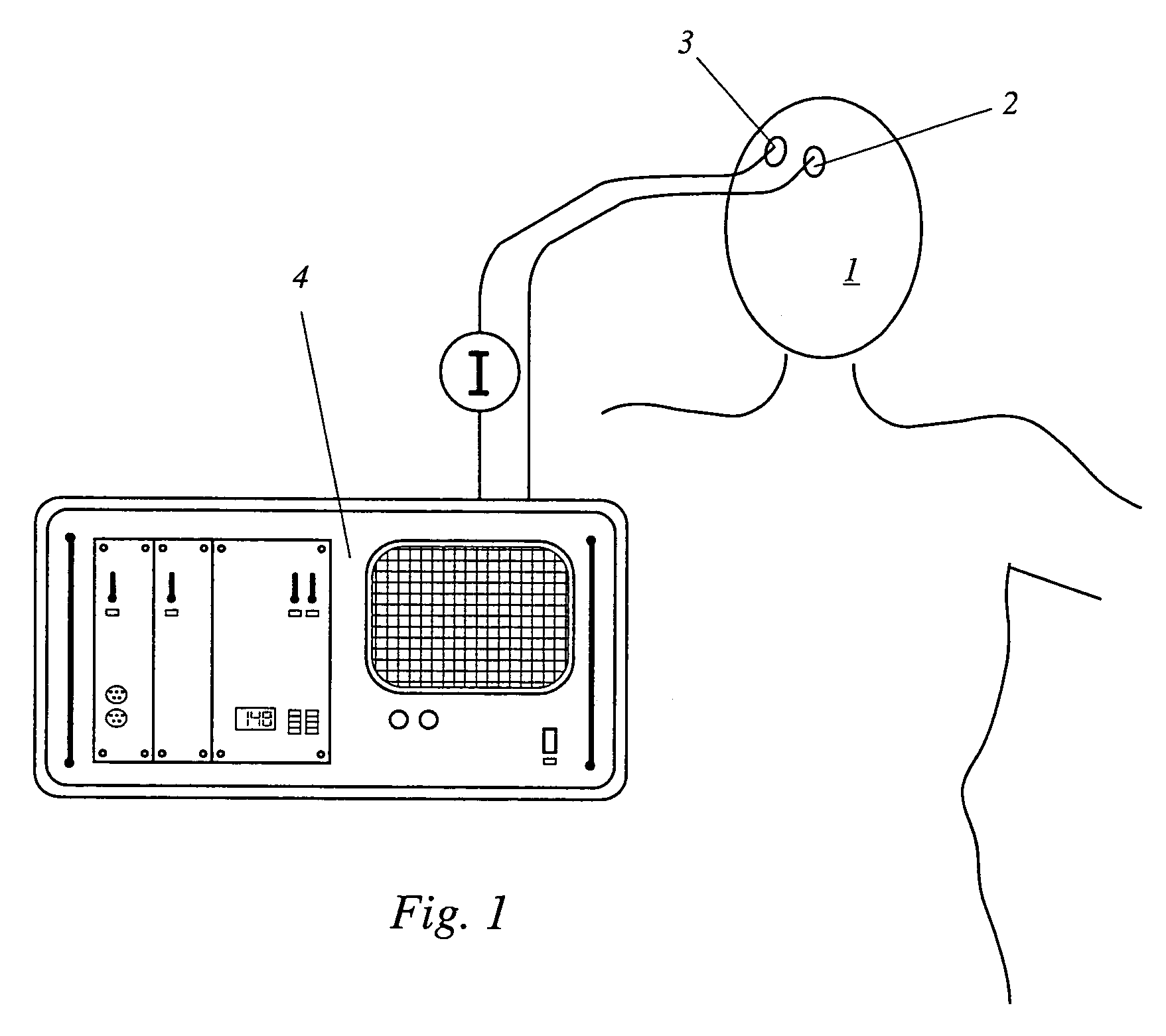
[0023]The device according to the invention shown schematically in FIG. 1 serves to determine the cerebral blood flow of a patient, for example an intensive-care patient in neurosurgery. A first optode 2 and a second optode 3 are attached to the head 1 of the patient, by means of an elastic band (not shown), at an optimized distance from one another. A radiation source (not shown) for emitting near infrared radiation into the cerebral tissue of the patient is arranged either in first optode 2 itself, or separately from it, for example in a common housing with the evaluation unit 4. Where the radiation source is separate from first optode 2, the near infrared radiation is passed by means of a light guide to first optode 2, where the radiation is emitted. The wavelength of the emitted radiation and the indicator used must be coordinated with one another. For the indicator usually used, indocyaningreen, a wavelength of about 805 nm (but definitely in the range between 780 and 910 nm) i...


PUM


Abstract

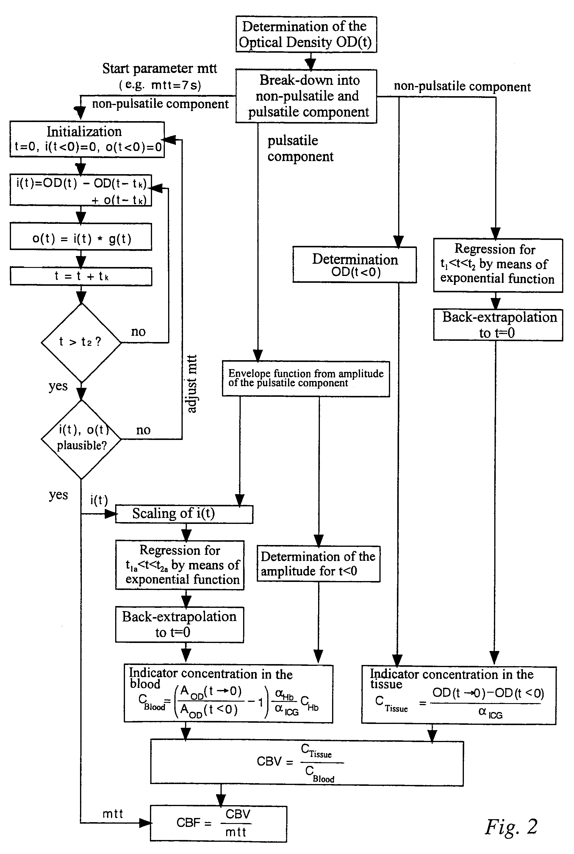
A device has a source for emitting near infrared radiation into cerebral tissue, a sensor for detecting radiation exiting from the tissue, and an evaluation unit which detects the exiting radiation as an input signal having pulsatile and non-pulsatile components and is programmed to determine the concentration of an injected indicator in the tissue from the non-pulsatile signal component, iteratively determine an inflow function characterizing cerebral blood flow by varying a mean transit time until reaching a stop criterion, determine indicator concentration relative to cerebral blood volume from the inflow function and the pulsatile signal component, calculate cerebral blood volume by dividing indicator concentration in the tissue by indicator concentration relative to cerebral blood volume, calculate cerebral blood flow by dividing the cerebral blood volume by the mean transit time when the stop criterion has been reached, and scale the inflow function using values determined from the pulsatile signal component.

Description

CROSS REFERENCE TO RELATED APPLICATIONS[0001]Applicants claim priority under 35 U.S.C. §119 of German Application No. 103 15 574.0 filed Apr. 5, 2003.BACKGROUND OF THE INVENTION[0002]1. Field of the Invention[0003]The present invention relates to a device for measuring the blood flow in an organ, using an injected indicator.[0004]2. The Prior Art[0005]Devices for measuring the blood flow in an organ, particularly the cerebral blood flow (CBF), using an injected, essentially inert indicator, have been known for a long time, and some of them are in clinical use. Devices based on older methods, such as the xenon dilution technique (Obrist, W. D., Thompson, H. K., Wang, H. S., and Wilkinson, W. F. 1975, Regional cerebral blood flow estimated by 133 xenon inhalation. Stroke 6: 245-256), are often difficult to implement, in technical terms, and prove to be time-consuming to use. Recently, technologies that use near-infrared spectroscopy (NIRS) in combination with indocyaningreen (ICG) as ...

Claims


Application Information

Patent Timeline
Patent Type & Authority Patents(United States)
IPC IPC(8): A61B6/00A61B5/026A61B5/0275
CPCA61B5/0275A61B5/0261A61B5/4064
Inventor KELLER, EMANUELANADLER, ANDREASNIEDERER, PETER
Owner UNIV ZURICH
Learn More