
Method for determining a path along a biological object with a lumen

a biological object and path technology, applied in the field of path determination along a biological object with a lumen, can solve the problems of high cost of paths partially or wholly outside the vessel, high computation overall cost, and execution times of several tens of seconds, so as to reduce the time taken to identify, less computation, and the effect of reducing the tim

Active Publication Date: 2008-05-27
TOSHIBA MEDICAL VISUALIZATION SYST EURO
View PDF22 Cites 27 Cited by
  • Summary
  • Abstract
  • Description
  • Claims
  • Application Information

AI Technical Summary

Benefits of technology

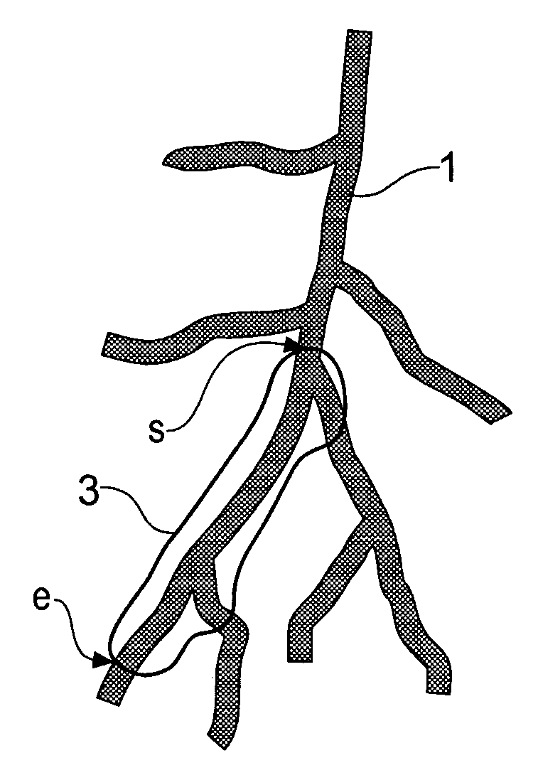
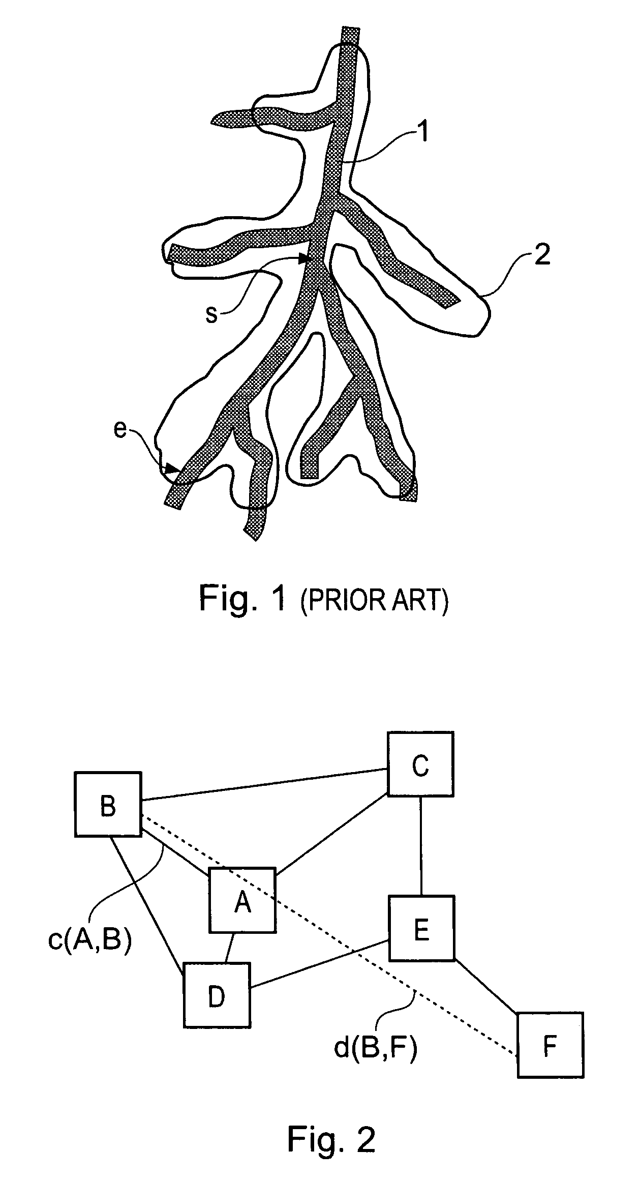
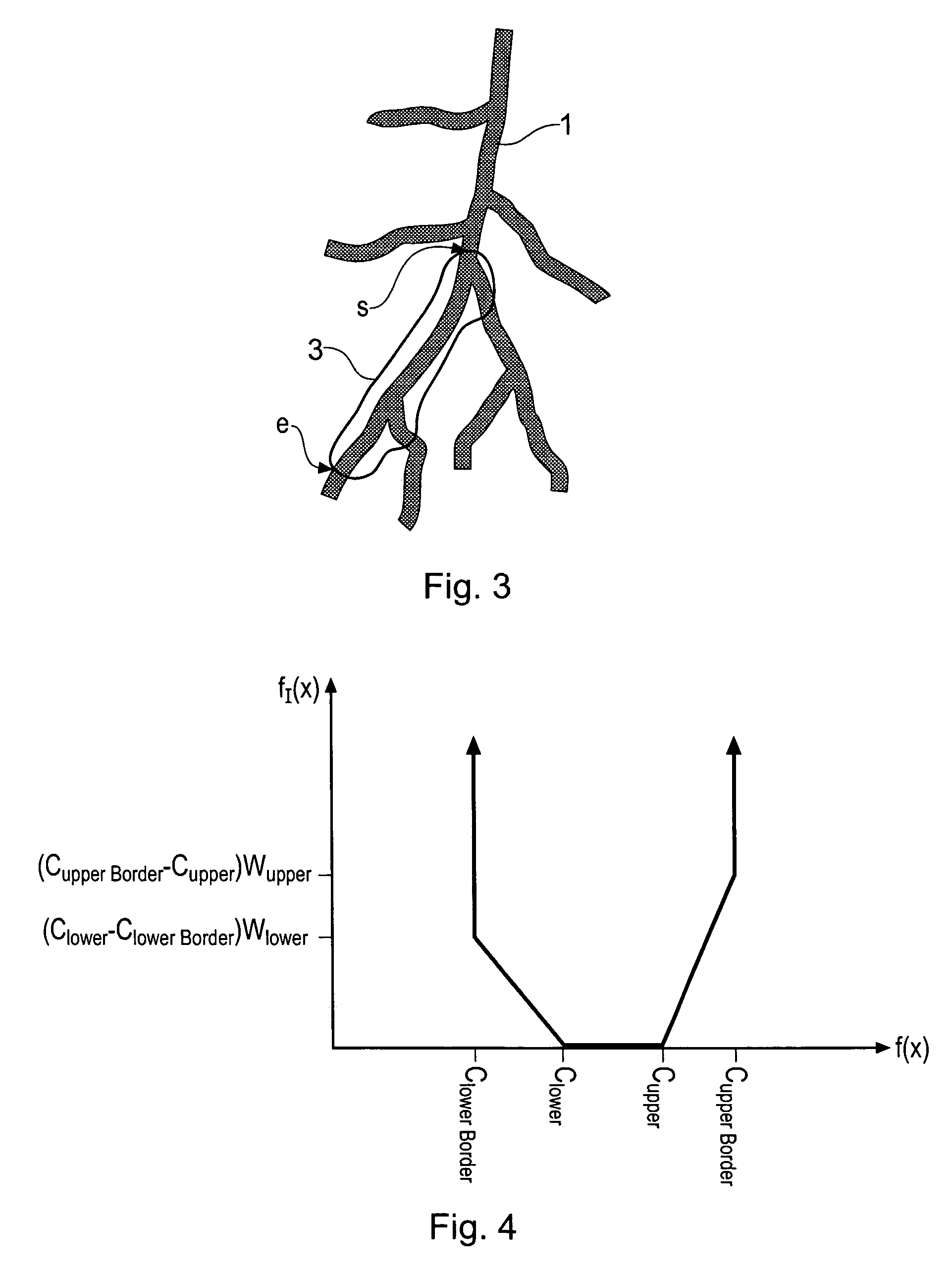
[0008]Accordingly, a first aspect of the present invention is directed to a computer-implemented method of determining a path along a three-dimensional object represented by a three-dimensional volume data set, the data set comprising a plurality of voxels each having a value indicating a characteristic of material represented by the voxel and arranged in a three-dimensional array, the method comprising: selecting a path start voxel s and a path end voxel e from voxels lying within the object; and determining a path from the start voxel s to the end voxel e via one or more intermediate voxels n lying within the object by using a minimum cost path finding algorithm to select the intermediate voxels n, where the algorithm prioritizes voxels for possible selection using a priority function that depends on a cost function that defines costs for paths between voxels, and on the Euclidean distance from a voxel to the end voxel e such that a smaller Euclidean distance gives a greater contribution to priority for that voxel, and the Euclidean distance is weighted by a constant k which is greater than zero.
[0009]This path finding method thus operates by taking account of the closeness of a candidate voxel to the end point of the path. The effect of this is to greatly reduce the time taken to identify a path between desired start and end points because far less computation is wasted exploring paths that lead away from the end point, compared to path finding techniques such as that proposed by Kanitsar [12, 13]. This advantageous development has been made possible by the recognition by the inventors of the present invention that a patient image three-dimensional volume data set composed of an array of voxels can be treated as a Euclidean network. In a preferred embodiment, the minimum cost path finding algorithm is a modified version of Dijkstra's algorithm.
[0010]Various embodiments of the method can be utilised by selecting the value of the constant k in the priority function. The following values of k are meaningful in the context that the cost of a path between two voxels is greater than or equal to the distance between those two voxels in any chosen units. For example, one may chose 0<k≦1, or k=1. In the case of a modified Dijkstra's algorithm, this regime allows the algorithm to find the lowest cost path along the object between the selected start and end voxels but much more quickly than if the Euclidean distance is not used in the prioritising (in other words, if k=0, which is Dijkstra's standard algorithm). The reduction in processing time is highly valuable for many patient image processing applications, such as stent planning and stenosis sizing, in which a real-time user experience is appreciated and possibly demanded.

Problems solved by technology

The cost function is the distance between the two voxels weighted by factors that give a low cost to paths lying within the vessel and a high cost to paths partially or wholly outside the vessel.
However, a disadvantage of Dijkstra's algorithm is that the computation overall can be very slow.
In the context of lumen path finding for applications such as stent planning and stenosis sizing, this is a serious problem that can result in execution times of several tens of seconds in many cases, which users tend to find unacceptably slow.

Method used


Image

  • Method for determining a path along a biological object with a lumen
  • Method for determining a path along a biological object with a lumen
  • Method for determining a path along a biological object with a lumen

Examples


first embodiment

[0041]The present inventors have recognised that this problem with Dijkstra's algorithm can be addressed by a modification. They have appreciated that a medical image three-dimensional volume data set can be considered to be a Euclidean network. In a Euclidean network, pluralities of nodes are connected by branches of known distance. In addition, the absolute spatial position of every node is known, so that the Euclidean distance (“as the crow flies” or shortest straight line distance) between any pair of nodes can be calculated. In the case of the three-dimensional volume data set, the voxels are regularly spaced in an array with known separations between the columns and rows of voxels (depending on the resolution of the imaging system used and any subsequent data processing) so that the Euclidean distance between any pair of voxels (not only adjacent voxels) is known. Incorporating this information into Dijkstra's algorithm provides a significant improvement to the path finding.

[0...

third embodiment

[0060]For some lumen path finding applications, in particular stent planning, stenosis sizing and virtual endoscopy, it is necessary or preferable to find the centerline path. This is the path that runs approximately along the geometric longitudinal axis of the lumen. For stent planning and stenosis sizing, a centerline enables accurate measurements to be taken from the anatomical region. In virtual endoscopy, it is desirable that the virtual camera follows the centerline path to avoid the impression to the user that the camera is about to collide with the object wall, and also to give the best possible view of the environment within the lumen to increase the chance that abnormalities are detected.

[0061]It is highly unlikely for any practical start and end point for a path in a lumen of interest that the shortest or lowest cost path will be the centerline path, particularly in a highly curved and / or branched network, where the shortest path will tend to hug the object wall around co...


PUM


Abstract

A path between specified start and end voxels along a biological object with a lumen, such as a vessel, within a patient image three-dimensional volume data set comprising an array of voxels of varying value is identified using an algorithm that works outwards from the start voxel to identify paths of low cost via intermediate voxels. The intermediate voxels are queued for further expansion of the path using a priority function comprising the sum of the cost of the path already found from the start voxel to the intermediate voxel and the Euclidean distance from the intermediate voxel to the end voxel. A cost function that depends on the voxel density is used to bias the algorithm towards paths inside the object. The number of iterations of the voxel required to find a path from the start to the end voxel, and hence the time taken, can be significantly reduced by scaling the Euclidean distance by a constant. Usefully, the constant is greater than 1, such as between 1.5 and 2.

Description

BACKGROUND OF THE INVENTION[0001]The present invention relates to a method for determining a path, which may be a centerline path, along a biological object having a lumen that is included in a three-dimensional volume data set representing a biological object.[0002]Patient imaging methods such as computer-assisted tomography (CT), magnetic resonance imaging (MRI), ultrasound and positron-emission tomography (PET) generate large three-dimensional volume data sets representing all or part of a patient's body. These volume data sets are highly detailed and allow complex studies of the body to be made. Advanced image processing techniques enhance the utility of the visualization of these data sets. For example, a physician may use a three-dimensional data set of an artery to determine the extent of stenosis or to plan the stenting of an aneurysm. This requires accurate identification of the centerline path along the lumen and measurements associated with the anatomy and physiology. Oth...

Claims


Application Information

Patent Timeline
Patent Type & Authority Patents(United States)
IPC IPC(8): G06T17/00A61B6/00G06K9/00
CPCG06T7/0083G06T7/60G06T2207/10072G06T2207/20101G06T2207/20136G06T2207/30101G06T7/12G06T7/181
Inventor POOLE, IAN
Owner TOSHIBA MEDICAL VISUALIZATION SYST EURO
Learn More