Method for diagnosis and treatment of haemophilia A patients with an inhibitor
a technology of inhibitors and haemophilia, applied in the field of diagnosis and medical treatment, can solve the problems of inadequate factor viii replacement therapy, ineffective treatment in all cases, and side effects of porcine factor viii administration, and achieve the effect of reducing the activity of factor viii inducing antibodies
- Summary
- Abstract
- Description
- Claims
- Application Information
AI Technical Summary
Benefits of technology
Problems solved by technology
Method used
Image
Examples
example 1
ization of Anti-factor VIII Antibodies in Patient's Plasma
[0059]Anti-factor VIII antibodies present in the plasma of a patient with acquired haemophilia were characterized by immunoprecipitation and neutralization experiments. The construction of recombinant factor VIII fragments corresponding to the A2, A3-C1-C2 and C2-domain of factor VIII has been described perviously (Fijnvandraat et al. 1997. Blood 89: 4371-4377; Fijnvandraat et al. 1998. Blood 91: 2347-2352). These recombinant factor VIII fragments were metabolically labelled with [35S]-methionine and subsequently used for the detection of anti-factor VIII antibodies by immunoprecipitation using methods that have been described previously (Fijnvandraat et al. 1998. Blood 91: 2347-2352). Reactivity with both metabolically labeled A2, A3-C1-C2 and C2 domain was observed (data not shown). This indicates that at least two classes of antibodies directed against factor VIII were present in the plasma of the patient. To determine the...
example 2
ion of an IgG4 Specific Library
[0060]Peripheral blood lymphocytes were isolated from a blood sample of a patient with acquired haemophilia. The titre of the inhibitor was 1250 BU / ml. RNA was isolated from the lymphocytes using RNAzol (WAK Chemie, Germany) according to the instructions of the manufacturer. RNA was transcribed into cDNA employing random hexamer primers (Gibco, Breda, The Netherlands). Since, most of the anti-factor VIII antibodies described in Example 1 were of subclass IgG4, DNA fragments corresponding to the heavy chain of immunoglobulins of subclass IgG4 were amplified using the following set of oligonucleotide primers:
[0061]
(SEQ. ID. NO:1)conIgG1-45′ CTTGTCCACCTTGGTGTTGCTGGG 3′(SEQ. ID. NO:2)huIgG45′ ACGTTGCAGGTGTAGGTCTTC 3′(SEQ. ID. NO:3)huVH1aback5′ CAGGTGCAGCTGGTGCAGTCTGG 3′(SEQ. ID. NO:4)huVH2aback5′ CAGGTCAACTTAAGGGAGTCTGG 3′(SEQ. ID. NO:5)huVH3aback5′ GAGGTGCAGCTGGTGGAGTCTGG 3′(SEQ. ID. NO:6)huVH4aback5′ GAGGTGCAGCTGTTGCAGTCGGG 3′(SEQ. ID. NO:7)huVH5aback5′ ...
example 3
of Factor VIII Specific Antibodies
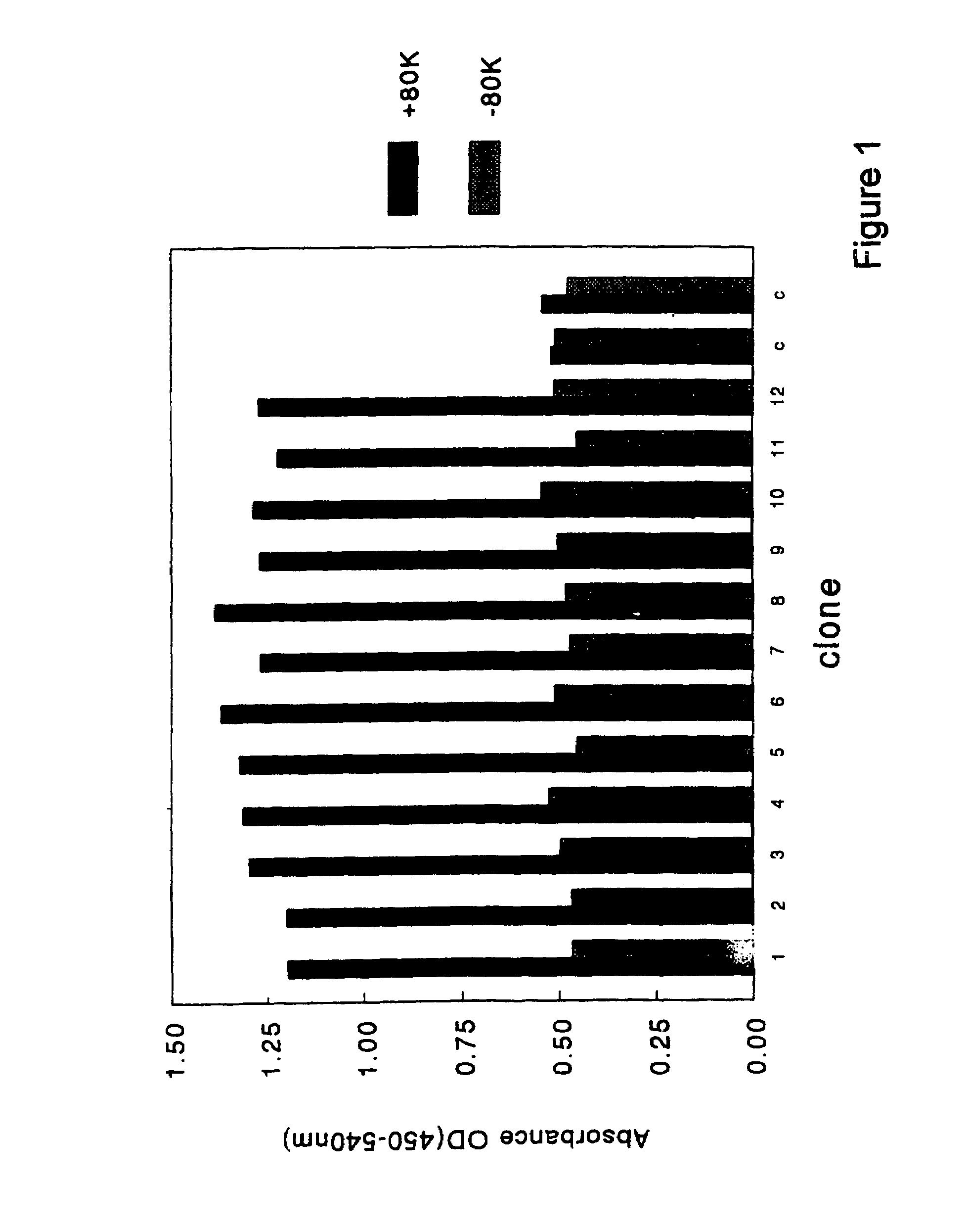
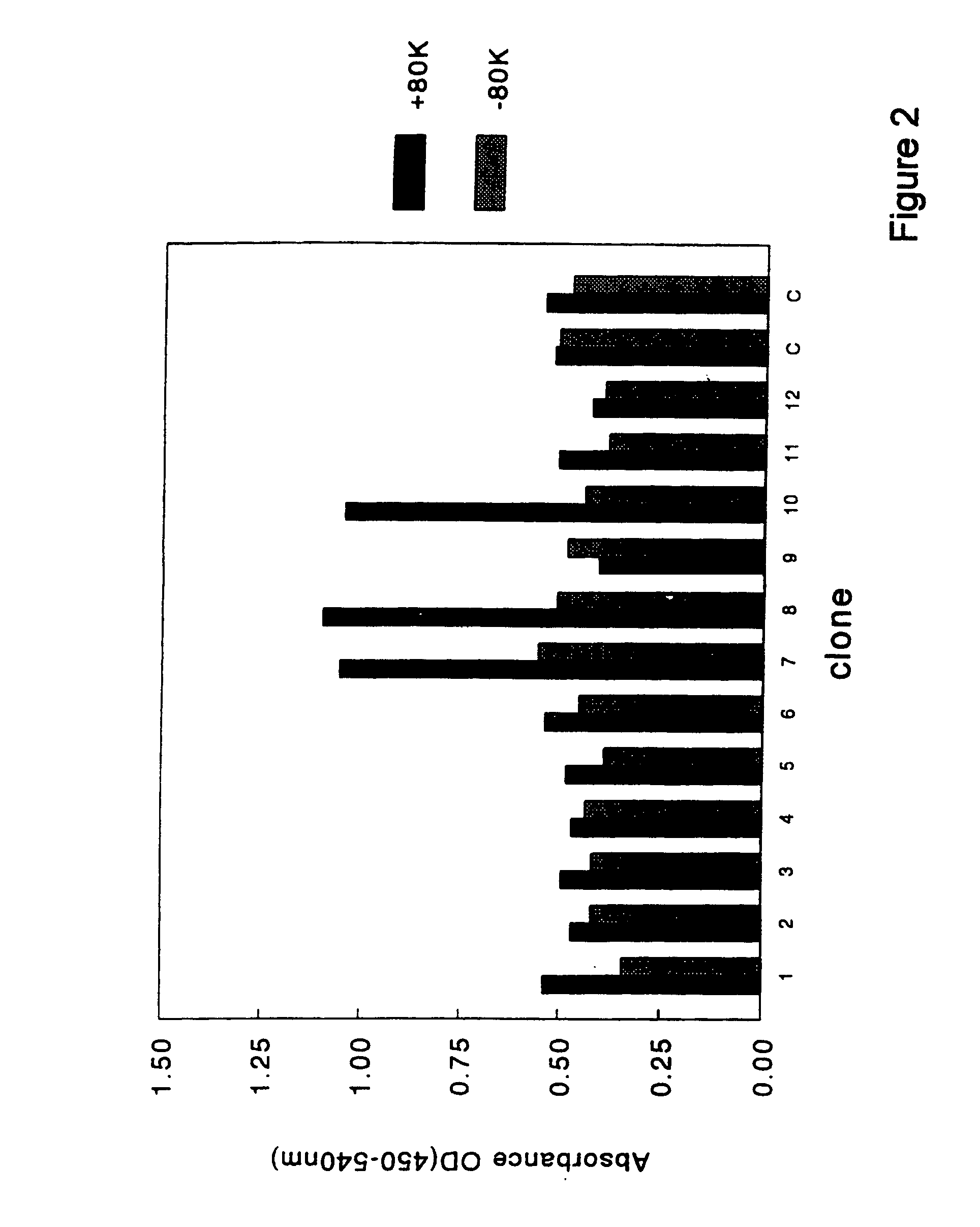
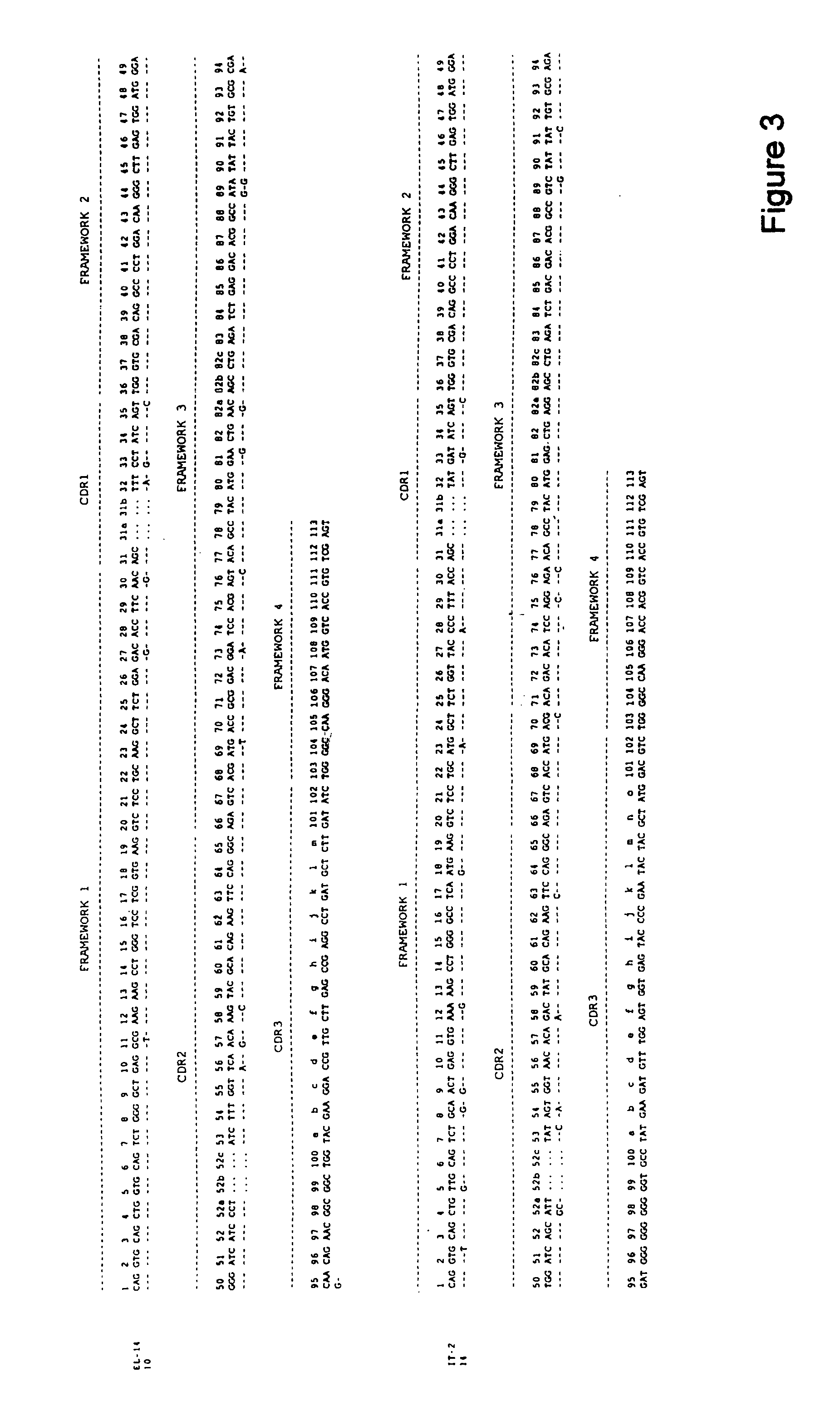
[0063]Selection of clones that encoded antibody fragments (scFvs) with factor VIII specificity was performed as outlined below. Glycerol stocks were plated onto 2TY plates that contained ampicillin (100 μg / ml) and 1% glucose. Colonies were grown overnight and scraped the next day and dissolved in 2TY supplemented with 100 μg / ml ampicillin and 1% glucose. These cells were diluted in 2TY supplemented with ampicillin (100 μg / ml) and 1% glucose till a final optical density (OD) of 0.3 (measured at 600 nm). Cells were grown at 37° C. till an OD of 0.5. Subsequently, 1 ml of culture was diluted 10 times in 2TY with ampicillin (100 μg / ml) and 1% glucose. Next, 20 μl of helper phage was added (VCSM13; 1×10¹¹ pfu / ml) and the mixture was incubated for 45 minutes at 37° C. without shaking. Then, cells were incubated at 37° C. with shaking at 150 rpm for another 45 minutes. The cells were spun down at low speed and resuspended in 100 ml of 2TY supplemented with...
PUM
 Abstract
Description
Claims
Application Information
 - R&D
- Intellectual Property
- Life Sciences
- Materials
- Tech Scout
- Unparalleled Data Quality
- Higher Quality Content
- 60% Fewer Hallucinations
Browse by: Latest US Patents, China's latest patents, Technical Efficacy Thesaurus, Application Domain, Technology Topic, Popular Technical Reports.
© 2025 PatSnap. All rights reserved.Legal|Privacy policy|Modern Slavery Act Transparency Statement|Sitemap|About US| Contact US: help@patsnap.com


