Identification and characterization of reflectin proteins from squid reflective tissues
a technology of reflectin and protein, applied in the field of proteins, can solve the problems of some or all of the incident light reflecting, and the composition of cephalopod reflector platelets has never been definitively characterized
- Summary
- Abstract
- Description
- Claims
- Application Information
AI Technical Summary
Benefits of technology
Problems solved by technology
Method used
Image
Examples
example 1
Isolation of Reflectin Proteins from E. scolopes
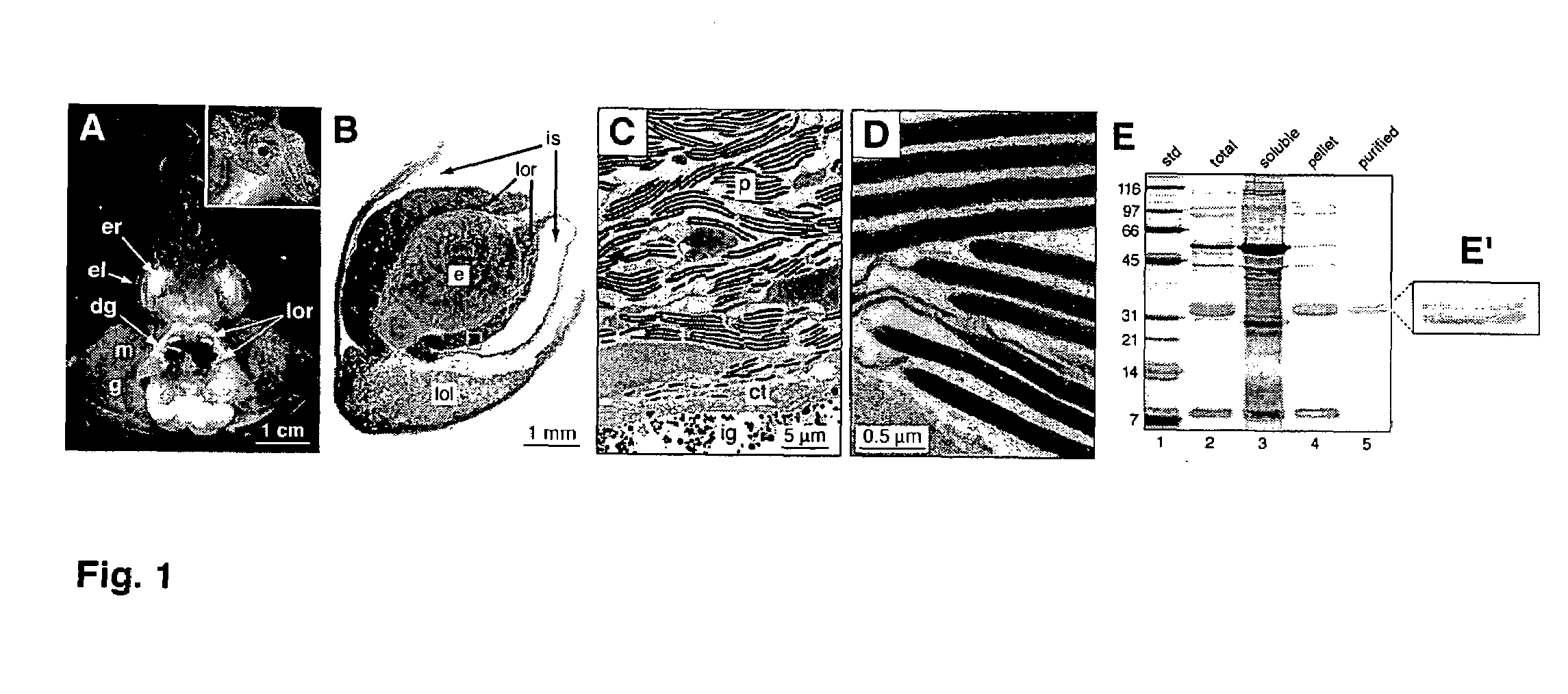
[0054]To enrich for the reflecting, the light organ reflector (LOR) was first homogenized in 50 mM sodium phosphate buffer, pH 7.4, with 0.1 M NaCl (PBS) in a ground glass homogenizer on ice to extract the aqueous soluble fraction. The total homogenate was centrifuged at 20,800×g for 15 min at 4° C. The resulting supernatant was removed. The pelleted material was then washed by repeatedly resuspending it in PBS and centrifuging the resuspension at 20,800×g for 15 min at 4° C. to re-pellet the aqueous-insoluble material. The resulting washed pellet was resuspended in 2% SDS in PBS to extract the SDS-soluble fraction that contained the reflecting. The suspension was then centrifuged, as described above, and the supernatant retained for analyses.
[0055]Sodium dodecyl sulfate-polyacrylamide gel electrophoresis (SDS-PAGE) was carried out on a Bio-Rad Mini-Protean II electrophoresis system (Bio-Rad, Hercules, Calif.) under standard SDS-PAGE ...
example 2
Tissue Localization of Reflectins
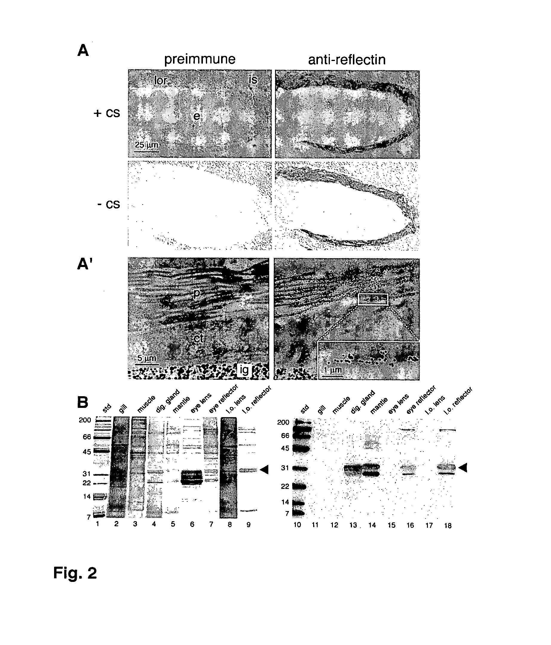
[0058]To localize the reflecting within the LOR, polyclonal antibodies were generated against gel-purified reflectin proteins (FIG. 1E, lane 5) and used in immunocytochemical and immunoblot analyses.
[0059]To generate material for antibody production, the reflectin proteins were purified directly from the SDS-PAGE gel of the LOR proteins. Briefly, the 2% SDS-soluble fraction of light organ reflectors from seven adult animals was applied to a stacking gel without wells. The portion of the gel containing the reflectins was excised from the gel and homogenized in 2% SDS in PBS. The resulting slurry was transferred to a Microcon-100 spin filter (Millipore Corp., Bedford, Mass.) and centrifuged at 3000×g for 10 min at room temperature. An aliquot of the filtrate was resolved on SDS-PAGE, that indicated that the desired proteins had been isolated. The remaining filtrate protein was used in the production of polyclonal antibodies (Covance Research Products, ...
example 3
Sequencing and Analysis of Reflectins Genes
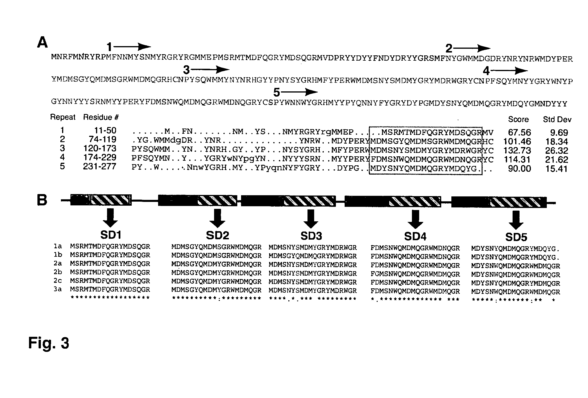
[0063]The sequences of three tryptic peptides (FIG. 3A) from tryptic digestion of gel-purified reflectins (FIG. 1E, lane 5) were used to identify reflectin cDNAs from predicted translations of E. scolopes cDNA and EST library clones as described below. The tryptic peptides had ambiguities in them as expressed by an “X” in the following sequences: SMFNYGWMMDGDR (SEQ ID NO:31), EGYYPNYSYGR (SEQ ID NO:32), and YFDMSNWQMDMQGR (SEQ ID NO:33).
[0064]Protein extracts from light organs were subjected to SDS-PAGE. Reflectin bands were excised from the gel and subjected to trypsin digestion. The resulting tryptic peptides were sequenced by mass spectrometry (Harvard Microchemistry Facility, Cambridge, Mass.). The amino acid sequences of three tryptic peptides were used to screen predicted translations of sequences from cDNA pools constructed from the light organs of juvenile animals (SEQ ID NOs: 31-33). One partial sequence was obtained from this pool...
PUM
| Property | Measurement | Unit |
|---|---|---|
| pH | aaaaa | aaaaa |
| pH | aaaaa | aaaaa |
| molecular weights | aaaaa | aaaaa |
Abstract
Description
Claims
Application Information
 - R&D
- Intellectual Property
- Life Sciences
- Materials
- Tech Scout
- Unparalleled Data Quality
- Higher Quality Content
- 60% Fewer Hallucinations
Browse by: Latest US Patents, China's latest patents, Technical Efficacy Thesaurus, Application Domain, Technology Topic, Popular Technical Reports.
© 2025 PatSnap. All rights reserved.Legal|Privacy policy|Modern Slavery Act Transparency Statement|Sitemap|About US| Contact US: help@patsnap.com


