
Computer-aided detection methods in volumetric imagery

a computer-aided detection and volumetric imaging technology, applied in image enhancement, diagnostic recording/measuring, instruments, etc., can solve the problems of difficult main task of identifying polyps, time-consuming virtual colonoscopy interpretation, and patient discomfor

Active Publication Date: 2007-06-26
ICAD INC
View PDF9 Cites 54 Cited by
  • Summary
  • Abstract
  • Description
  • Claims
  • Application Information

AI Technical Summary

Problems solved by technology

However, current screening methods of fiberoptic colonoscopy, flexible sigmoidoscopy, fecal occult blood testing and barium enema examination carry varying amounts of risk and may cause considerable discomfort to the patient, and therefore are not widely accepted by the general population.
Because of the large number of images that need to be examined in 2D views (300-700 images / patient) and the relatively long time it takes to do an endoluminal “fly-through,” virtual colonoscopy interpretation can be quite time-consuming, typically ranging from 15 to 40 minutes when performed by experts in abdominal imaging.
Additionally, the main task of identifying polyps can be challenging.

Method used


Image

  • Computer-aided detection methods in volumetric imagery
  • Computer-aided detection methods in volumetric imagery
  • Computer-aided detection methods in volumetric imagery

Examples


example outputs

[0029]To demonstrate the utility of the present invention, FIGS. 5-7 show a set of detector outputs on transverse slices from CT imagery. The outline of the colon is divided into three objects on these slices. Separate objects appear in these transverse views due to vertical trajectories of the colon. Circles represent detections found by the method of the present invention and rectangles represent areas of visually identified polyps. The utility of the present invention is confirmed by noting detections within regions identified as polyps.


PUM


Abstract

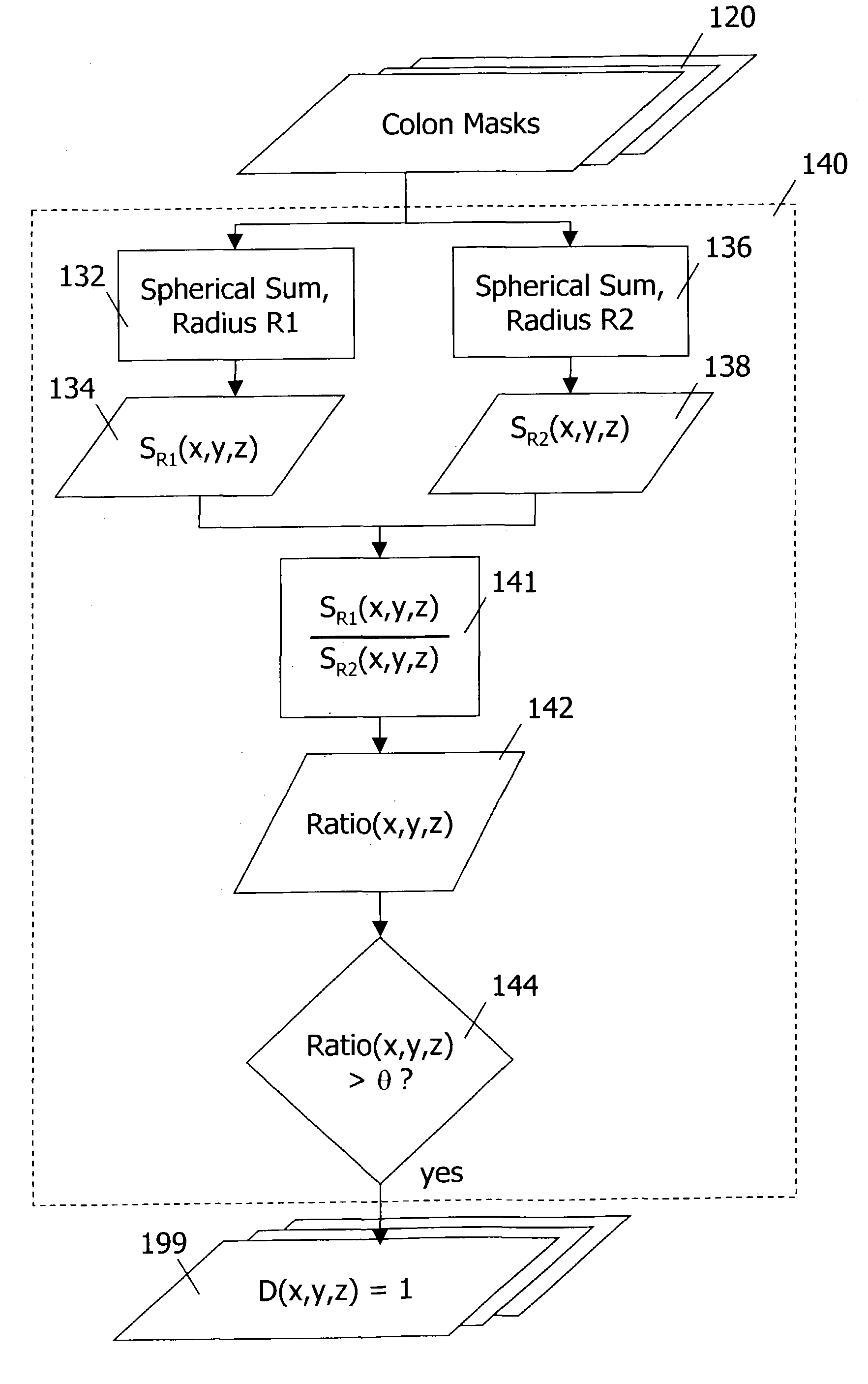
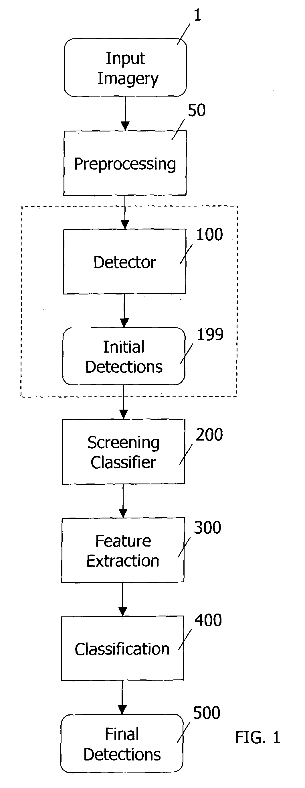
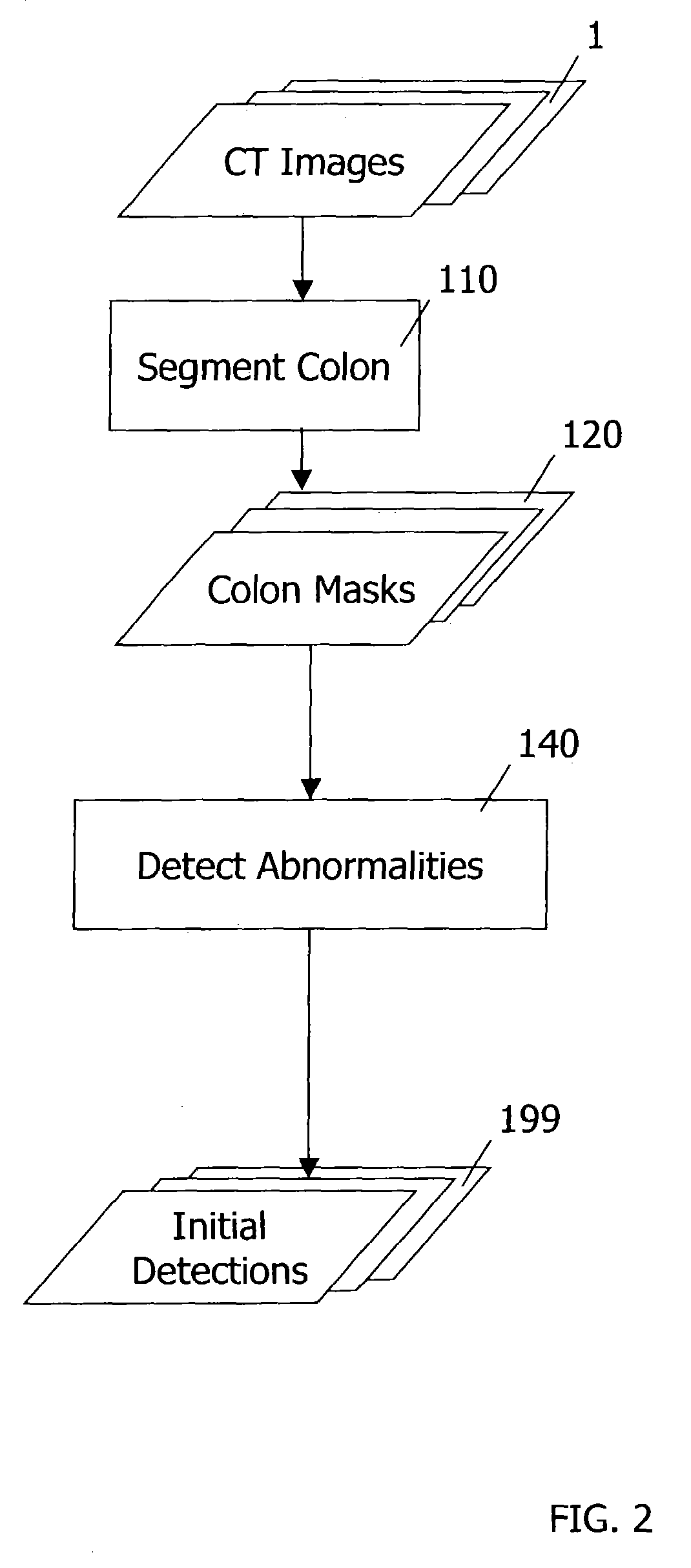
This invention provides a method for detecting abnormalities in medical images, particularly for the detection of polyps in the colon from computed tomography imagery. Specifically a set of colon masks is input to a detector where summations over spherical volumes with two different radii are computed. Abnormalities are detected based on the ratio of spherical summation values computed for every pixel location.

Description

CROSS-REFERENCE TO RELATED APPLICATIONS[0001]This application claims the benefit of U.S. Provisional Application No. 60 / 391,120, filed Jun. 24, 2002, incorporated herein by reference.BACKGROUND OF THE INVENTION[0002]1. Field of the Invention[0003]This invention relates to a method and system for computer-aided detection (CAD) of volumetric images, specifically for detection of polyps indicative of colon cancer in computed tomography imagery.[0004]2. Discussion of Background[0005]Colorectal cancer is the second leading cause of cancer death in the United States. Each year approximately 150,000 people are diagnosed with colon cancer and 56,000 people die from it. Colonoscopic removal of identifiable polyps can considerably decrease the mortality. However, current screening methods of fiberoptic colonoscopy, flexible sigmoidoscopy, fecal occult blood testing and barium enema examination carry varying amounts of risk and may cause considerable discomfort to the patient, and therefore ar...

Claims


Application Information

Patent Timeline
Patent Type & Authority Patents(United States)
IPC IPC(8): G06K9/00G06V10/40
CPCG06K9/46G06T7/0012G06T2207/30028G06V10/40
Inventor GURCAN, METIN N.
Owner ICAD INC
Learn More