
Molecular modification assays

a molecular modification and assay technology, applied in the field of molecular modification assays, can solve the problems of short-term safety hazards for the assay operator, long-term storage and disposal problems, and inability to obtain suitable antibodies for many substrates and kinases, and achieve the effects of decreasing rotational mobility and increasing luminescence polarization of the protein

Inactive Publication Date: 2006-07-04
MOLECULAR DEVICES
View PDF353 Cites 91 Cited by
  • Summary
  • Abstract
  • Description
  • Claims
  • Application Information

AI Technical Summary

Benefits of technology

[0030]Errors in the signal transduction and regulation pathways described above can cause cancer and other diseases. Indeed, a primary cause of cancer is a mutation that makes a stimulatory gene product hyperactive, converting a proto-oncogene into an oncogene. The primary classes of known proto-oncogenes include the following cell-signaling proteins: (1) growth-factor receptors acting via tyrosine kinases, (2) GTP binding proteins, (3) membrane / cytoskeleton-associated tyrosine kinases, (4) cytoplasmic tyrosine kinases, (5) steroid-type growth-factor receptors, and (6) S / T kinases. Consequently, cell-signaling proteins have become important subjects of research and drug development.
[0031]Assays that determine the presence and / or activity of cell-signaling components are important tools in life sciences research, including high-throughput screening. Unfortunately, current assays have a number of shortcomings.
[0032]The presence and activity of kinases, for example, can be determined using assays capable of detecting phosphorylated amino acids. In a standard kinase assay, radioactive ATP and an appropriate protein substrate are added to a sample. If the sample includes kinases, radioactive phosphate groups will be transferred from the radioactive ATP to the protein substrate. The presence and activity of kinases can be determined by assaying the amount of radioactive protein substrate, for example, using heterogeneous methods such as a filter plate that involve separating the protein substrate and radioactive ATP, or homogeneous methods such as a scintillation proximity assay for detecting radioactive decay. Unfortunately, both approaches involve radioactivity, presenting a short-term safety hazard for the assay operator and a long-term storage and disposal problem.
[0033]In an alternative kinase assay, ATP, a luminescent protein, and an antibody against a phosphorylated form of the luminescent protein are added to a sample. If the sample includes kinases, the kinases will transfer phosphate groups from the ATP to the protein, the antibody will bind to the phosphorylated protein, and the luminescence polarization of the protein will increase (because its rotational mobility will decrease). Unfortunately, the binding of antibodies is very target specific, so that in general a different antibody will be needed for each substrate (depending on the sequence of the substrate, including whether a tyrosine, serine, or threonine is to be phosphorylated). This shortcoming is especially significant for serines and threonines. Thus, a different antibody may be needed for each of the many kinases, depending on the polypeptide sequences of the corresponding substrates. Yet, suitable antibodies may be unavailable for many substrates and kinases, especially for poorly studied or previously unstudied kinases, or take several months or more to prepare.
[0034]Significantly, assays for other cell-signaling components (such as phosphatases, phosphodiesterases, and / or cyclases) may have similar shortcomings, such as the use of radioactive reagents, if the assays exist at all. Moreover, these assays may have slow time courses and unstable endpoints that require precise timing of assay readouts. Thus, there is a need for improved assays for detecting enzyme activity, and in particular the presence and activity of cell-signaling components.
[0035]The invention provides assays for detecting molecular modifications such as phosphate modifications and the presence and / or activity of enzymes and other agents involved in facilitating or otherwise regulating such modifications.

Problems solved by technology

Errors in the signal transduction and regulation pathways described above can cause cancer and other diseases.
Unfortunately, current assays have a number of shortcomings.
Unfortunately, both approaches involve radioactivity, presenting a short-term safety hazard for the assay operator and a long-term storage and disposal problem.
Unfortunately, the binding of antibodies is very target specific, so that in general a different antibody will be needed for each substrate (depending on the sequence of the substrate, including whether a tyrosine, serine, or threonine is to be phosphorylated).
Yet, suitable antibodies may be unavailable for many substrates and kinases, especially for poorly studied or previously unstudied kinases, or take several months or more to prepare.
Moreover, these assays may have slow time courses and unstable endpoints that require precise timing of assay readouts.

Method used


Image

  • Molecular modification assays
  • Molecular modification assays
  • Molecular modification assays

Examples


example 1

[0084]This example describes a macromolecular trapping system for use in luminescence polarization and / or energy transfer assays, among others, in accordance with aspects of the invention. In this system, Ru2+ is entrapped in small (˜20-30 kDa) synthetic polymer macromolecules (MM), which are obtained from PreSens Precision Sensing (Neuburg / Donau, Germany). These macromolecules are relatively hydrophilic, with carboxyl groups on their surfaces for activation. The MM with the entrapped Ru2+ is used as a support to immobilize tricationic metal cations, including Fe3+ and Ga3+. Specifically, the chelator imidodiacetic (IDA) acid is linked to the MM using the secondary amine group of IDA and a carboxyl group on the MM. Afterwards, the MM-IDA is incubated with either FeCl3 or GaCl3. The FeCl3 quenches the luminescence of Ru2+, whereas the GaCl3 does not. The macromolecule loaded with Fe3+ or Ga3+ is denoted MM-Fe or MM-Ga, respectively.

[0085]The macromolecular trapping system may be used...

example 2

[0088]This example describes assays for the presence, activity, substrates, and / or products of kinases in accordance with aspects of the invention. Similar assays may be used to analyze phosphatases in which the substrates and products of the kinase reaction become the products and substrates of the phosphatase reaction, respectively.

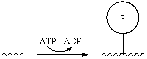
[0089]Kinases catalyze the addition of phosphate groups to appropriate substrates, as shown below:

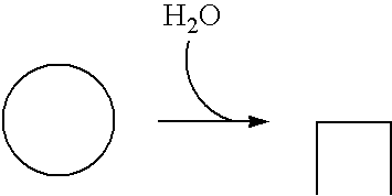
Thus, the presence and / or activity of a kinase may be detected by a decrease in the concentration of a nonphosphorylated (e.g., polypeptide) substrate and / or by an increase in the concentration of a corresponding phosphorylated product, among others. (The presence and / or activity of a phosphatase may be detected similarly by a decrease in the concentration of a phosphorylated substrate and / or an increase in the concentration of a nonphosphorylated product.) The invention provides among others kinase assays that involve contacting a sample containing a cand...

example 3

[0090]This example describes experiments to characterize binding between MM-Ga and a fluorescein-labeled di-phosphotyrosine 15-amino-acid peptide tracer denoted tyrosine kinase 1 (TK-1) tracer. These experiments show the utility of the MM-Ga system for detection of phosphorylated tyrosine and the presence and / or activity of tyrosine kinases and phosphatases.

[0091]FIG. 8 shows the effects of incubating 10 nM TK-1 tracer with different concentrations of MM-GA (total volume=50 μL; incubation time=60 min). These experiments show that the maximum polarization change is more than 200 mP, at least when the MM-Ga and TK-1 tracer are incubated in MES buffer (0.1M MES, pH 5.5, 1.0 M NaCl). This polarization change is at least sufficient for most polarization assays.

[0092]FIG. 9 shows a dose-response curve for TK-1 calibrator, with 10 nM TK-1 tracer and 1.6 nM (estimated) MM-Ga. The TK-1 calibrator is the same as the TK-1 tracer, without a fluorescein label. The bound / total ratio is calculated...


PUM

PropertyMeasurementUnit
volumesaaaaaaaaaa
pHaaaaaaaaaa
pHaaaaaaaaaa

Abstract

Assays for detecting molecular modifications such as phosphate modifications and the presence and / or activity of enzymes and other agents involved in facilitating or otherwise regulating such modifications.

Description

CROSS-REFERENCE[0001]This application is based upon and claims benefit under 35 U.S.C. §119(e) of the following U.S. provisional patent applications, which are incorporated herein by reference: Ser. Nos. 60 / 200,594, filed Apr. 28, 2000; Ser. No. 60 / 223,642, filed Aug. 8, 2000; and Ser. No. 60 / 241,032, filed Oct. 17, 2000.[0002]This application is a continuation of and claims benefit under 35 U.S.C. §120 of the following patent applications, which are incorporated herein by reference: PCT Patent Application Ser. No. PCT / US00 / 16025, filed Jun. 9, 2000; and is a continuation of U.S. patent application Ser. No. 09 / 596,444, filed Jun. 19, 2000.[0003]This application incorporates by reference the following U.S. patents: U.S. Pat. No. 5,843,378, issued Dec. 1, 1998; U.S. Pat. No. 6,965,381, issued Oct. 12, 1999; U.S. Pat. No. 6,071,748, issued Jun. 6, 2000; and U.S. Pat. No. 6,097,025, issued Aug. 1, 2000.[0004]This application also incorporates by reference the following U.S. patent appli...

Claims


Application Information

Patent Timeline
Patent Type & Authority Patents(United States)
IPC IPC(8): G01N21/75B01L3/00C07K7/06C07K7/08C12Q1/48G01N33/53G01N33/542G01N33/566G01N33/58G01N35/00G01N35/02G01N35/04G01N35/10
CPCB01L3/50853C07K7/08C12Q1/485G01N33/5308G01N33/542G01N33/566G01N33/582G01N35/028C07K7/06Y10S436/805G01N35/1011G01N35/1074G01N2035/00237G01N2035/0405G01N2035/0425G01N2035/0462G01N2333/9121Y10S436/80
Inventor HUANG, WEIHOEKSTRA, MERL F.SPORTSMAN, J. RICHARDTERPETSCHNIG, EWALD A.
Owner MOLECULAR DEVICES
Learn More