
Methods and apparatus for detecting structural, perfusion, and functional abnormalities

a technology of functional abnormalities and methods, applied in the field of computed tomographic (ct) imaging, can solve the problems of not providing quantitative image values in conventional ct, other beam hardening artifacts may be difficult to correct, and other beam hardening artifacts such as metals and contrast agents, which are difficult to corr

Inactive Publication Date: 2006-06-06
GENERAL ELECTRIC CO
View PDF21 Cites 29 Cited by
  • Summary
  • Abstract
  • Description
  • Claims
  • Application Information

AI Technical Summary

Problems solved by technology

Some beam-hardening artifacts can be easily corrected, but other beam-hardening artifacts may be more difficult to correct.
However, beam hardening from materials other than water and bone, such as metals and contrast agents, may be difficult to correct.
In addition, even with the above described correction methods, conventional CT does not provide quantitative image values.
Another drawback of conventional CT is a lack of material characterization.
At least some state-of-the-art CT scanners currently available are limited to providing anatomical information.
For lung scans, images produced by such scanners exhibit a significant level of image artifacts and CT number inaccuracy.
These limitations prevent the utilization of the CT device for advanced diagnosis.

Method used


Image

  • Methods and apparatus for detecting structural, perfusion, and functional abnormalities
  • Methods and apparatus for detecting structural, perfusion, and functional abnormalities
  • Methods and apparatus for detecting structural, perfusion, and functional abnormalities

Examples


Embodiment Construction

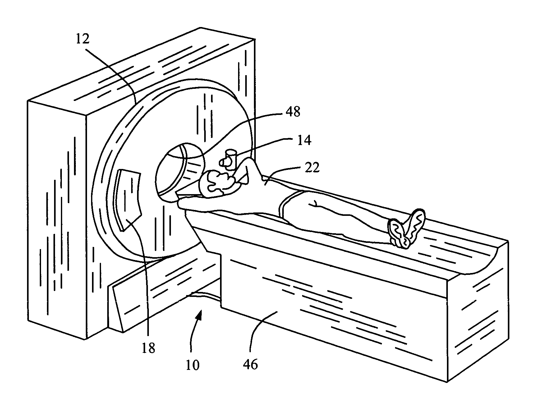
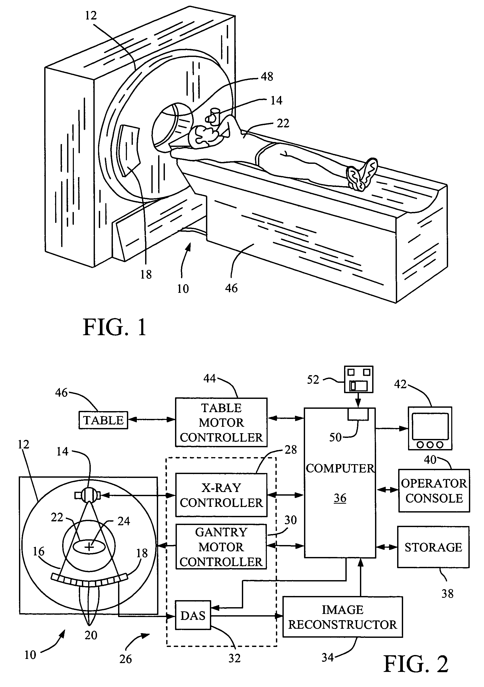
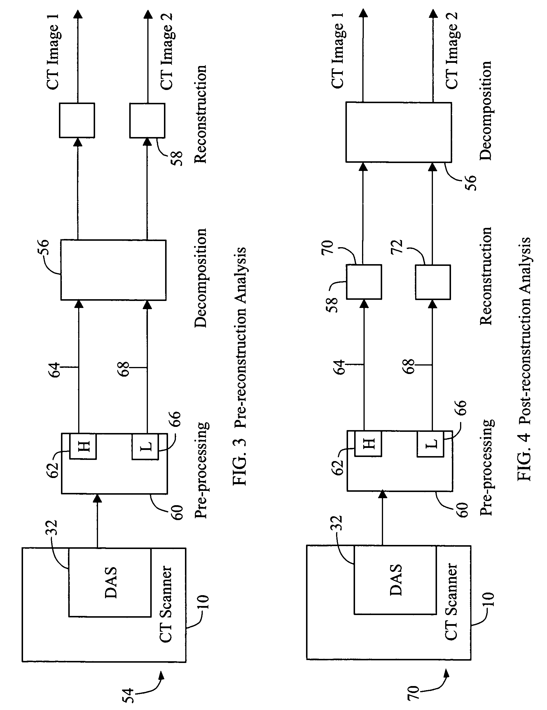
[0020]The methods and apparatus described herein address the detection and diagnosis of abnormalities in the lung regions of a patient by employing novel approaches that make use of basic properties of the x-ray and material interaction. For each ray trajectory, multiple measurements regarding different mean x-ray energies are acquired. As explained in greater detail below, when Basis Material Decomposition (BMD) and Compton and photoelectric decomposition are performed on these measurements, additional information is obtained that enables improved accuracy and characterization.

[0021]In some known CT imaging system configurations, an x-ray source projects a fan-shaped beam which is collimated to lie within an X-Y plane of a Cartesian coordinate system and generally referred to as an “imaging plane”. The x-ray beam passes through an object being imaged, such as a patient. The beam, after being attenuated by the object, impinges upon an array of radiation detectors. The intensity of t...


PUM

PropertyMeasurementUnit
CTaaaaaaaaaa
computed tomographyaaaaaaaaaa
energy resolutionaaaaaaaaaa

Abstract

A method for obtaining data includes scanning a lung of a patient with a Multi-Energy Computed Tomography (MECT) system to acquire data regarding a plurality of contrast agents.

Description

[0001]This application is a Continuation In Part (CIP) of U.S. patent application Ser. No. 10 / 306,467, filed Nov. 27, 2002, now U.S. Pat. No. 6,813,333 B1, issued Nov. 2, 2004, which is hereby incorporated by reference in its entirety.BACKGROUND OF THE INVENTION[0002]This invention relates to computed tomographic (CT) imaging, and more particularly to methods and apparatus for the detection and diagnosis of lung abnormalities.[0003]In spite of recent advancements in computed tomography (CT) technology, such as faster scanning speeds, larger coverage with multiple detector rows, and thinner slices, energy resolution is still a missing piece. Namely, wide x-ray photon energy spectrum from the x-ray source and the lack of energy resolution from CT detection systems preclude energy discrimination CT.[0004]X-ray attenuation through a given object is not a constant. Rather, the X-ray attenuation is strongly dependent on the x-ray photon energy. This physical phenomenon manifests itself in...

Claims


Application Information

Patent Timeline
Patent Type & Authority Patents(United States)
IPC IPC(8): G01N23/00A61B6/00A61B6/03
CPCA61B6/032A61B6/4035A61B6/405A61B6/504A61B6/4241A61B6/481A61B6/482A61B6/4085
Inventor PIACSEK, KELLY LYNNSABOL, JOHN MICHAEL
Owner GENERAL ELECTRIC CO
Learn More