Eureka AIR delivers breakthrough ideas for toughest innovation challenges, trusted by R&D personnel around the world.

Methods and devices using magnetic force to form an anastomosis between hollow bodies

a technology of hollow body and magnetic force, which is applied in the direction of prosthesis, surgical staples, blood vessels, etc., can solve the problems of high technical and time-consuming procedure of creating handsewn anastomosis, heart disease remains the leading cause of death, and manipulating the aorta puts patients at risk, so as to facilitate the anastomosis

Inactive Publication Date: 2005-08-23
MEDTRONIC INC
View PDF49 Cites 124 Cited by
  • Summary
  • Abstract
  • Description
  • Claims
  • Application Information

AI Technical Summary

Benefits of technology

The invention provides a method and system for forming an anastomosis between two hollow bodies in a patient's body using magnetism. The method involves positioning two securing components in the hollow bodies and using magnetic force to bring them into communication with each other. This allows the lumens of the hollow bodies to be placed in communication. The system includes the securing components, which produce a magnetic field to maintain their desired relative position. The technical effects of the invention include the ability to form an anastomosis without the need for surgery or external devices, and the potential for reduced risk of complications and recovery time for patients.

Problems solved by technology

Despite the considerable advances that have been realized in cardiology and cardiovascular surgery, heart disease remains the leading cause of death throughout much of the world.
Clamping or otherwise manipulating the aorta places the patient at risk in some cases due to the likelihood that such manipulation will release embolic material into the bloodstream.
Some challenges associated with this procedure include delivering and deploying the conduit in the patient's body, properly positioning the conduit with respect to the heart chamber and the target vessel, and obtaining beneficial flow characteristics through the conduit and the target vessel.
A particularly challenging task that must be performed during CABG procedures (as well as some ventricular bypass procedures) is suturing the conduit to one or more vessels.
The small diameter of the vessels involved (typically from 1 mm to 4 mm) makes creating a handsewn anastomosis a highly technical and time-consuming procedure.
The difficulty in forming the sutured anastomosis is exacerbated when access to the target vessel is restricted or limited, as in a minimally invasive or percutaneous procedure.
This problem also arises in non-cardiovascular applications that utilize handsewn anastomoses, for example, treating peripheral vascular disease or injury, creating AV (arteriovenous) shunts, etc.
While those in the art have proposed various anastomotic couplings intended to replace a sutured anastomosis, none has performed well enough to receive any significant level of acceptance in the field.
Many of the proposed couplings penetrate or damage the target vessel wall acutely or chronically, do not remain patent, fail to produce a fluid-tight seal between the conduit and vessel, or are simply too cumbersome and difficult to deliver or deploy.

Method used


Image

  • Methods and devices using magnetic force to form an anastomosis between hollow bodies
  • Methods and devices using magnetic force to form an anastomosis between hollow bodies
  • Methods and devices using magnetic force to form an anastomosis between hollow bodies

Examples


Embodiment Construction

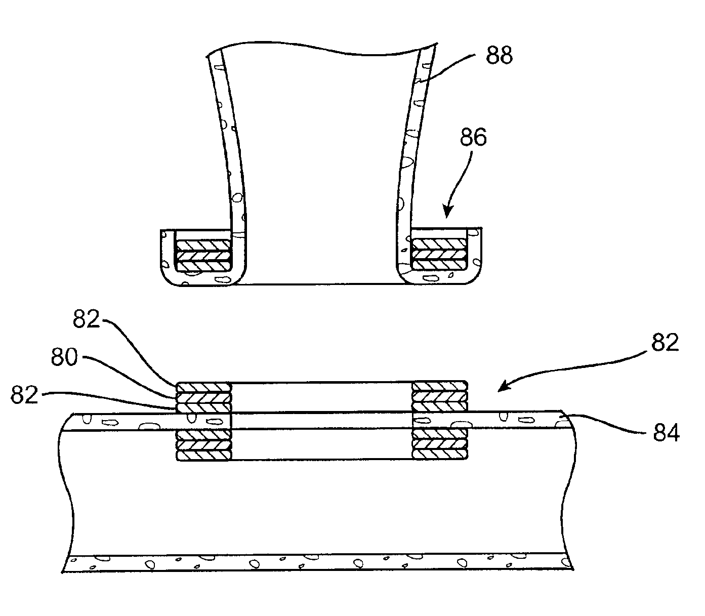
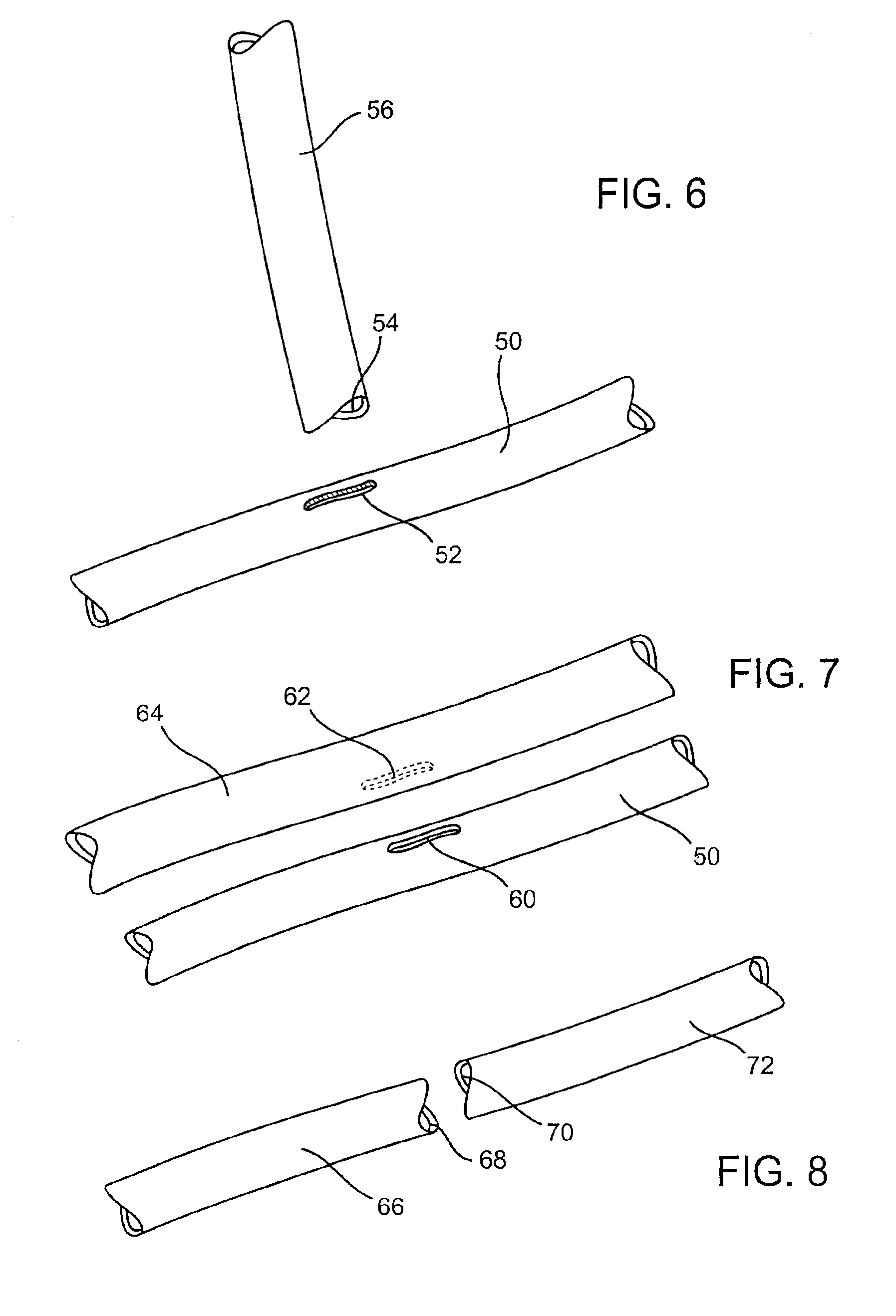
[0052]The present invention relates to methods and devices for forming an anastomosis between first and second (or additional) hollow bodies located in a patient's body, for example, a connection between a graft vessel and coronary or peripheral blood vessels, viscera, reproductive ducts, etc. The anastomosis places the hollow bodies, more specifically the lumens of the hollow bodies, in communication. In the case of blood-carrying bodies (or other hollow bodies that carry fluid) the anastomosis places the bodies in fluid communication. The hollow bodies being joined may comprise native or autologous vessels, vessels formed of synthetic material such as ePTFE, DACRON®, etc.

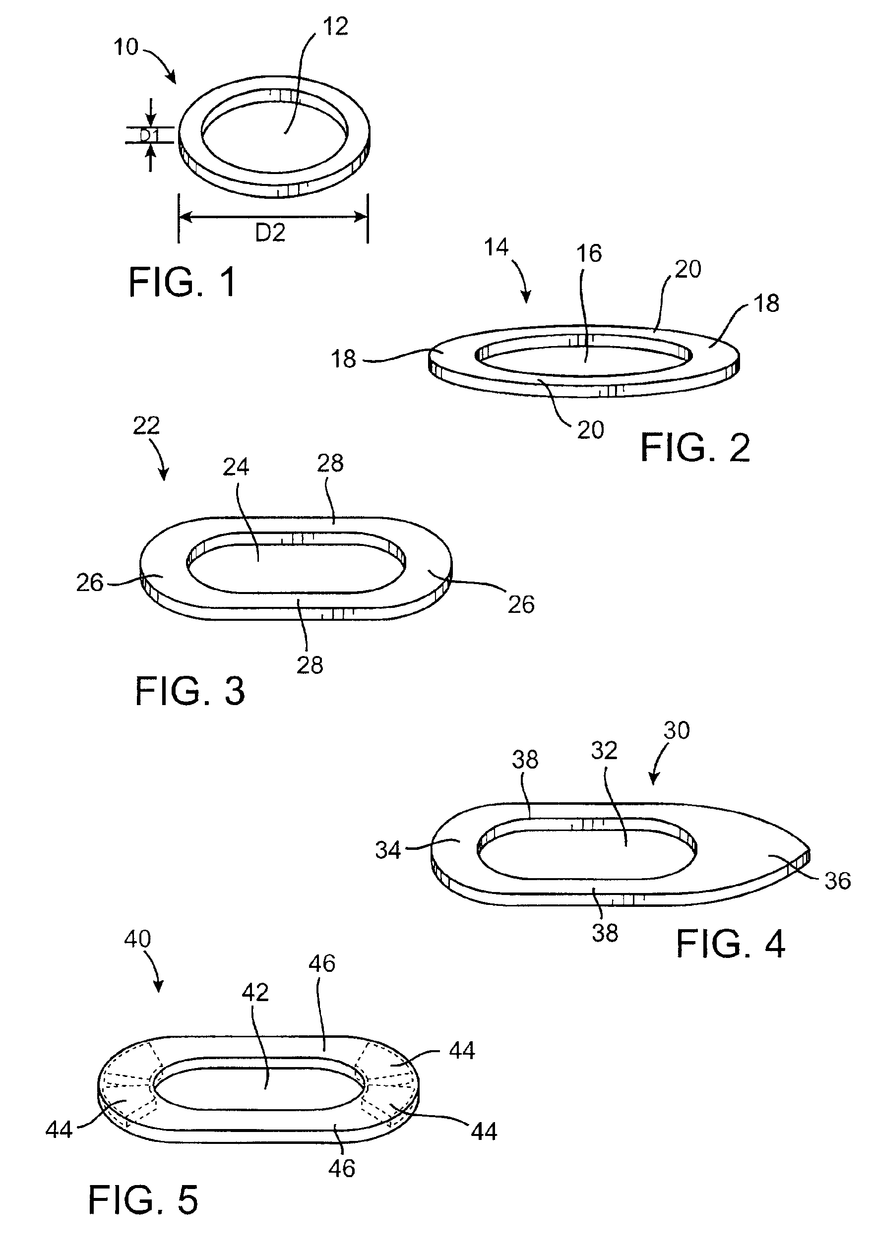
[0053]FIGS. 1-5 illustrate several exemplary embodiments of anastomotic securing components constructed according to the invention for use in forming an anastomosis between first and second hollow bodies. FIG. 1 shows a securing component 10 with an annular body and an opening 12 defined by the body. The component...


PUM

PropertyMeasurementUnit
diameteraaaaaaaaaa
thickaaaaaaaaaa
thickaaaaaaaaaa

Abstract

Methods and devices for forming an anastomosis between hollow bodies utilizes magnetic force to couple anastomotic securing components and create a fluid-tight connection between the lumens of the hollow bodies. End-to-side, side-to-side and end-to-end anastomoses can be created without using suture or any other type of mechanical fasteners, although any such attachment means may be used in conjunction with the magnetic attachment. The securing components have magnetic, ferromagnetic or electromagnetic properties and may include one or more materials, for example, magnetic and nonmagnetic materials arranged in a laminated structure. The system of anastomotic securing components may be used in many different applications including the treatment of cardiovascular disease, peripheral vascular disease, forming AV shunts for dialysis patients, etc., and may be sized and configured for forming an anastomosis to a specific hollow body, for example, a coronary artery or the aorta.

Description

CROSS-REFERENCE TO RELATED APPLICATION[0001]This application is a continuation of application Ser. No. 09 / 562,599, filed Apr. 29, 2000 U.S. Pat. No. 6,352,543. The complete disclosure of the aforementioned related U.S. patent application is hereby incorporated herein by reference for all purposes.BACKGROUND OF THE INVENTION[0002]1. Field of the Invention[0003]The invention relates to methods and devices for forming an anastomosis between two hollow bodies, and more particularly to forming an anastomosis using magnetic force.[0004]2. Description of Related Art[0005]Despite the considerable advances that have been realized in cardiology and cardiovascular surgery, heart disease remains the leading cause of death throughout much of the world. Coronary artery disease, or arteriosclerosis, is the single leading cause of death in the United States today. As a result, those in the cardiovascular field continue to search for new treatments and improvements to existing treatments.[0006]Coron...

Claims


Application Information

Patent Timeline
Patent Type & Authority Patents(United States)
IPC IPC(8): A61B17/11A61B17/03A61F2/00A61B17/00A61B17/04A61B17/064A61F2/02A61F2/06A61F2/24A61F2/94H01F7/02H01F41/02
CPCA61B17/0057A61B17/0643A61B17/11A61F2/2493A61F2/94A61B17/00491A61B17/0469A61B17/064A61B2017/00247A61B2017/00252A61B2017/00575A61B2017/00592A61B2017/00606A61B2017/00876A61B2017/1107A61B2017/1132A61B2017/1135A61B2017/1139A61B2018/00392A61F2/064A61F2/07A61F2002/30079A61F2210/009H01F7/0263H01F41/026A61F2210/0076
Inventor COLE, DAVID H.
Owner MEDTRONIC INC
Why Eureka
Eureka Blog
Learn More