Three dimensional protein mapping
a three-dimensional protein and mapping technology, applied in the field of multi-phase protein separation methods, can solve the problems of many limitations of traditional 2-d pages, and achieve the effect of efficient facilitation of protein sample transfer
- Summary
- Abstract
- Description
- Claims
- Application Information
AI Technical Summary
Benefits of technology
Problems solved by technology
Method used
Image
Examples
example 1
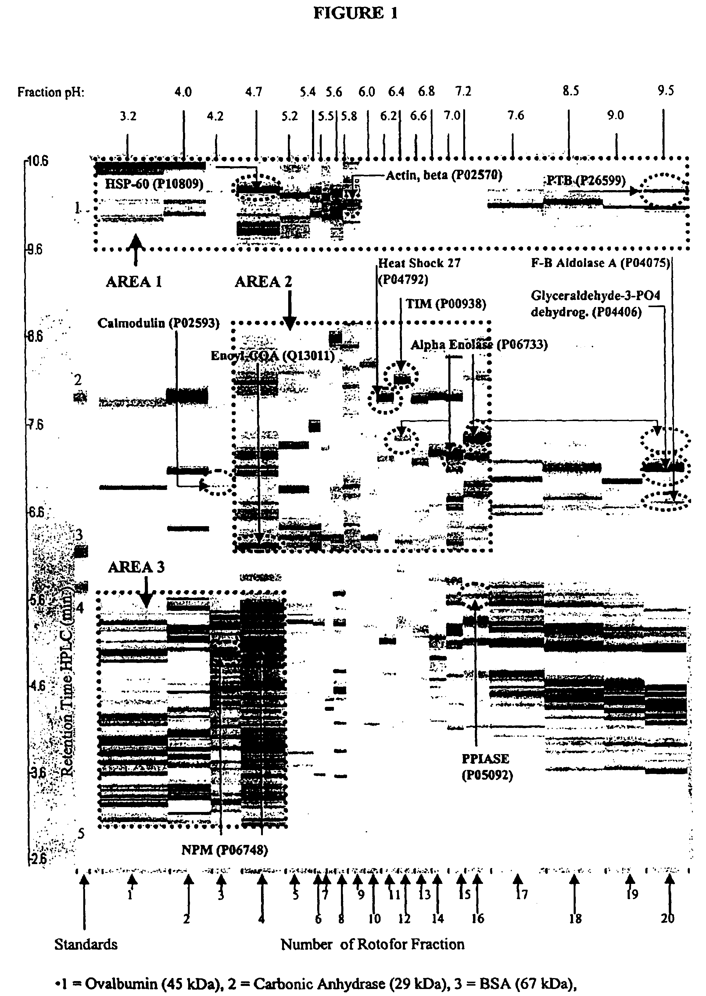
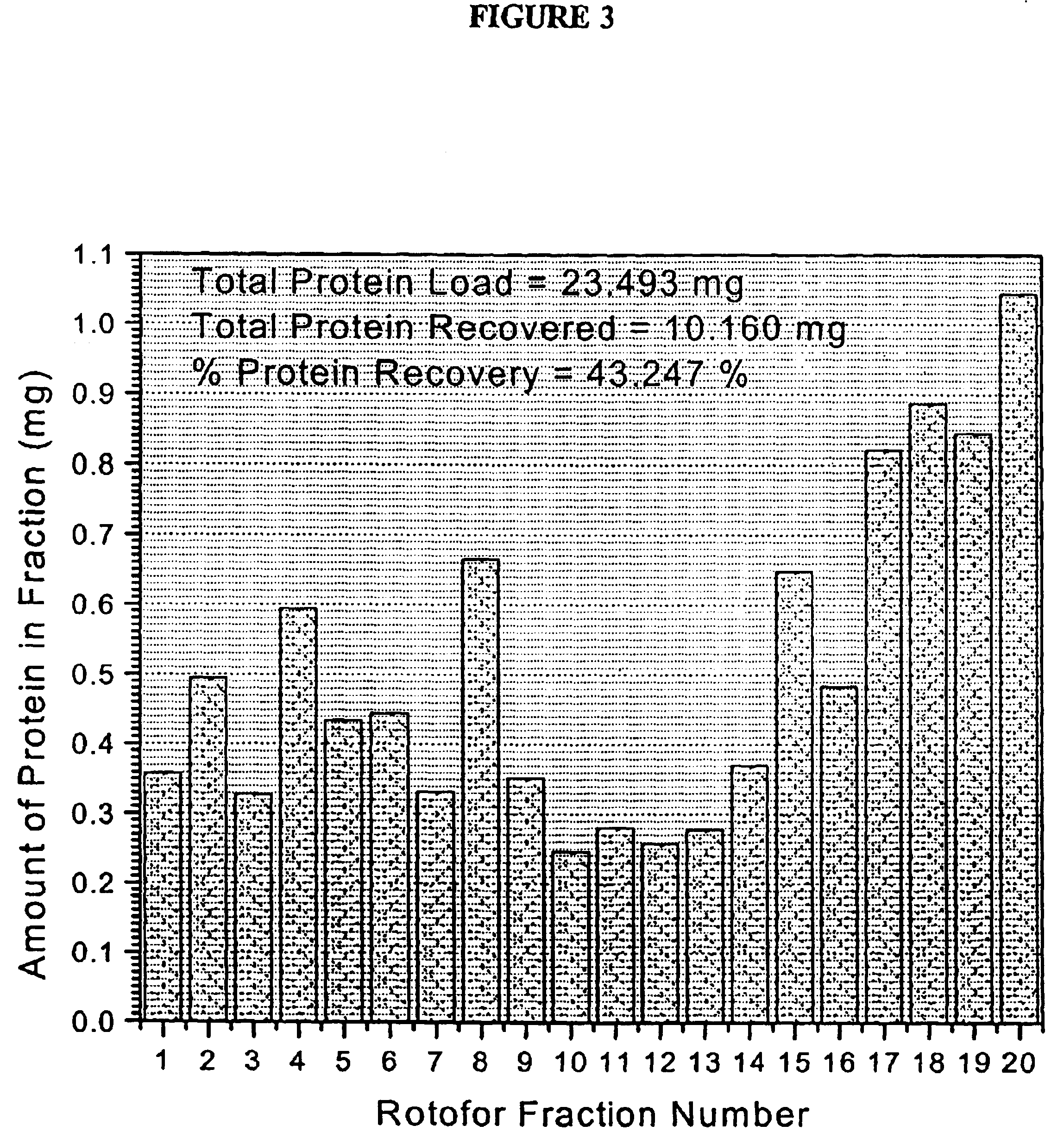
[0195]The human erythroleukemia (HEL) cell line was obtained from the Department of Pediatrics at The University of Michigan. HEL cells were cultured (7% CO2, 37° C.) in RPMI-1640 medium (Gibco) containing 4 mM glutamine, 2 mM pyruvate, 10% fetal bovine serum (Gibco), penicillin (100 units per mL), streptomycin (100 units per mL) and 250 mg of hygromycin (Sigma). The HEL cell pellets were washed in sterile PBS, and then stored at −80° C. The cell pellets were then re-suspended in 0.1% n-octyl β-D-galactopyranoside (OG)(Sigma) and 8 M urea (Sigma) and vortexed for 2 minutes to effect cell disruption and protein solubilization. The whole cell protein extract was then diluted to 55 mL with the Rotofor buffer and introduced into the Rotofor separation chamber (Biorad).
example 2
1-D Gel and SDS PAGE Separation
[0196]HEL cell proteins, resolved by Rotofor separation into discrete pI ranges, were further resolved according to their apparent molecular weight by SDS-PAGE. This procedure takes approximately 14 hours to complete. Samples of rotofor fractions were suspended in an equal volume of sample buffer (125 mM Tris (pH 6.8) containing 1% SDS, 10% glycerol, 1% dithiothreitol and bromophenol blue) and boiled for 5 min. They were then loaded onto 10% acrylamide gels. The samples were electrophoresed at 40 volts until the dye front reached the opposite end of the gel. The resolved proteins were visualized by silver staining. The gels were fixed overnight in 50% ethanol containing 5% glacial acetic acid, then washed successively (for 2 hours each) in 25% ethanol containing 5% glacial acetic acid, 5% glacial acetic acid, and 1% glacial acetic acid. The gels were impregnated with 0.2% silver nitrate for 25 min, and were developed in 3% sodium carbonate containing 0...
example 3
2-D PAGE
[0197]In order to prepare protein extracts from the HEL cells, the harvested cell pellets were lysed by addition of three volumes of solubilization buffer consisting of 8 M urea, 2% NP-40, 2% carrier ampholytes (pH 3.5 to 10), 2% β-mercaptoethanol and 10 mM PMSF, after which the buffer containing the cell extracts was transferred into microcentrifuge tubes and stored at −80° C. until use.
[0198]Extracts of the cultured HEL cells were separated in two dimensions as previously described by Chen et al (Chen et al., Rap. Comm. Mass Spec. 13:1907 [1999]) with some modifications as described below. Subsequent to cellular lysis in solubilization buffer, the cell lysates from approximately 2.5×10⁶ cells were applied to isoelectric focusing gels. Isoelectric focusing was conducted using pH 3.5 to 10 carrier ampholytes (Biorad) at 700 V for 16 h, followed by 1000 V for an additional 2 hours. The first dimension tube gel was soaked in a solution of 2 mg / mL of dithioerythritol (DTE) for ...
PUM
| Property | Measurement | Unit |
|---|---|---|
| mass | aaaaa | aaaaa |
| mass | aaaaa | aaaaa |
| temperature | aaaaa | aaaaa |
Abstract
Description
Claims
Application Information
 - R&D
- Intellectual Property
- Life Sciences
- Materials
- Tech Scout
- Unparalleled Data Quality
- Higher Quality Content
- 60% Fewer Hallucinations
Browse by: Latest US Patents, China's latest patents, Technical Efficacy Thesaurus, Application Domain, Technology Topic, Popular Technical Reports.
© 2025 PatSnap. All rights reserved.Legal|Privacy policy|Modern Slavery Act Transparency Statement|Sitemap|About US| Contact US: help@patsnap.com


