
Proteomic analysis

a proteome and proteome technology, applied in the field of proteome analysis, can solve the problems of insufficient knowledge of the genome sequence to explain biology and understand disease, methods that do not describe selectively detecting active versus inactive proteins within a sample, and methods that fail to report on enzyme activity

Inactive Publication Date: 2005-03-29
THE SCRIPPS RES INST
View PDF5 Cites 82 Cited by
  • Summary
  • Abstract
  • Description
  • Claims
  • Application Information

AI Technical Summary

Benefits of technology

Systems are provided for identifying portions of proteomes, where the proteome activity is determined for a plurality of active proteins and the effect of agents on the activity of such proteome portion. The system includes methods for identifying probes useful for reacting with active proteins, probes for reacting with active proteins and methods for identifying the

Problems solved by technology

Consequently, knowing the sequence of the genome is insufficient to explain biology and to understand disease.
These methods do not describe selectively detecting active versus inactive proteins within a sample.
Although current proteomic approaches, such as those described above, could theoretically provide information on the abundance of an enzyme, these methods fail to report on enzyme activity.
This is a key limitation because the activity of enzymes, and even other proteins, is often regulated by post-translational modification.
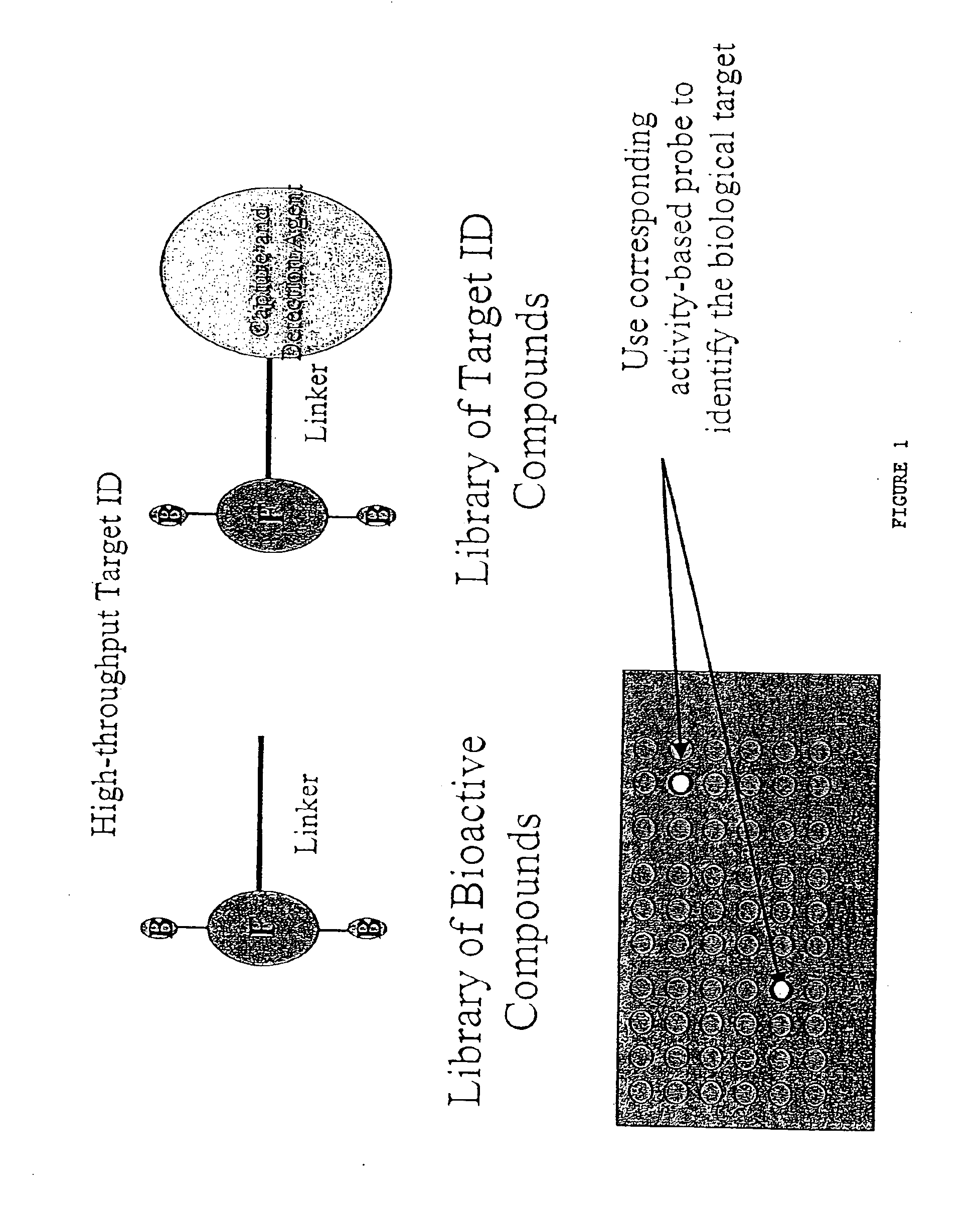
One of the inherent difficulties with the entire development process is that it is often difficult to identify the biological target molecule for lead compounds.
There is no simple way of determining what the biological target molecule is for lead compounds identified from such screens.
Nor, is there a way of knowing if multiple lead compounds interact with the same, or with different, biological target molecules.
Consequently, the identification of the biological target molecule relies on conventional fractionation and purification strategies, which are cumbersome, time consuming and expensive.
Moreover, without knowledge of the identity of the biological target molecule, and an understanding of its precise biochemical activity, one may be unable to devise assays to track its purification during these steps.
Consequently, the identity of such biological target molecules is often impossible to determine using current approaches.

Method used


Image

  • Proteomic analysis
  • Proteomic analysis
  • Proteomic analysis

Examples


example 1

Compound 1 is the starting material tetraethyleneoxy (3,6,9-oxa-1,11-diolundecane) as depicted in the flow chart in FIG. 8.

Compound 2. A solution of 1 (3.9 g, 20.0 mmol, 3.0 equiv) in DMF (8.0 mL) was treated with TBDMSCl (1.0 g, 6.64 mmol, 1.0 equiv) and imidazole (0.9 g, 13.3 mmol, 2.0 equiv) and the reaction mixture was stirred for 12 h at room temperature. The reaction mixture was then quenched with saturated aqueous NaHCO3 and partitioned between ethyl acetate (200 mL) and water (200 mL). The organic layer was washed with dried (Na2SO4) and concentrated under reduced pressure. Chromatography (SiO2, 5×15 cm, 50-100% ethyl acetate-hexanes) afforded 2 (1.1 g, 2.0 g theoretical, 55%) as a colorless oil: 1H NMR (CDCl3, 400 MHz) δ 3.8-3.5 (m, 16H, CH2OR), 0.88 (s, 9H, CH3C), 0.0 (s, 6H, CH3Si).

Compound 3. A solution of 2 (0.61 g, 2.0 mmol, 1.0 equiv) in benzene (15 mL, 0.13 M) was treated sequentially with PPh3 (2.6 g, 10.0 mmol, 5 equiv), 12 (2.3 g, 9.0 mmol, 4.5 equiv), and imidazo...

example 2

Preparation of FP-biotin

FP-Biotin was prepared as described by Liu et al. (Proc. Natl. Acad. Sci. 96(26):14694, 1999) and in U.S. Ser. Nos. 60 / 195,954 and 60 / 212,891, herein incorporated by reference in their entirety. 1-Iodo-10-undecene (3). A solution of 2 (3.4 g, 10.5 mmol, 1.0 equiv) in acetone (21 mL, 0.5 M) was treated with NaI (3.2 g, 21 mmol, 2.0 equiv) and the reaction mixture was stirred at reflux for 2 h, producing a yellow-orange solution. The reaction mixture was then partitioned between ethyl acetate (200 mL) and water (200 mL). The organic layer was washed sequentially with saturated aqueous Na2S2O3 (100 mL) and saturated aqueous NaCl (100 mL), dried (Na2SO4), and concentrated under reduced pressure. Chromatography (SiO2, 5×15 cm, 1-2% ethyl acetate-hexanes) afforded 3 (2.3 g, 2.9 g theoretical, 78%) as a colorless oil: 1H NMR (CDCl3, 250 MHz) δ 5.95-5.75 (m, 1H, RCH═CH2), 5.03-4.90 (m, 2H, RCH═CH2), 3.16 (t, J=7.0 Hz, 2H, CH2I), 2.02 (m, 2H, CH2CH═CH2), 1.80 (p, J=6....

example 3

Molecular Characterization of FP-biotin Reactive Proteins

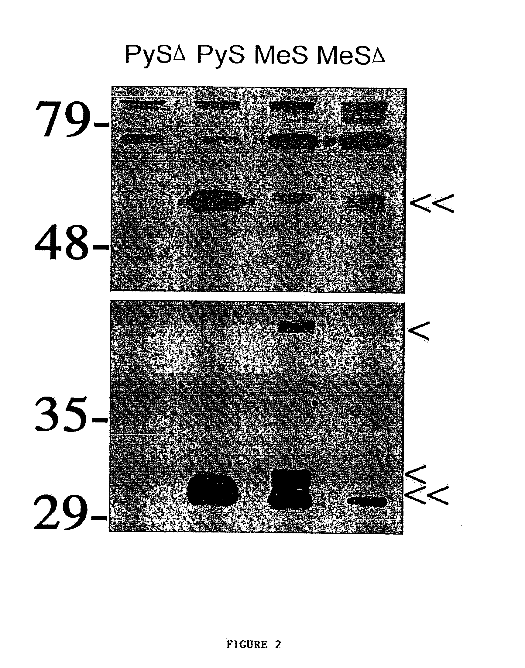
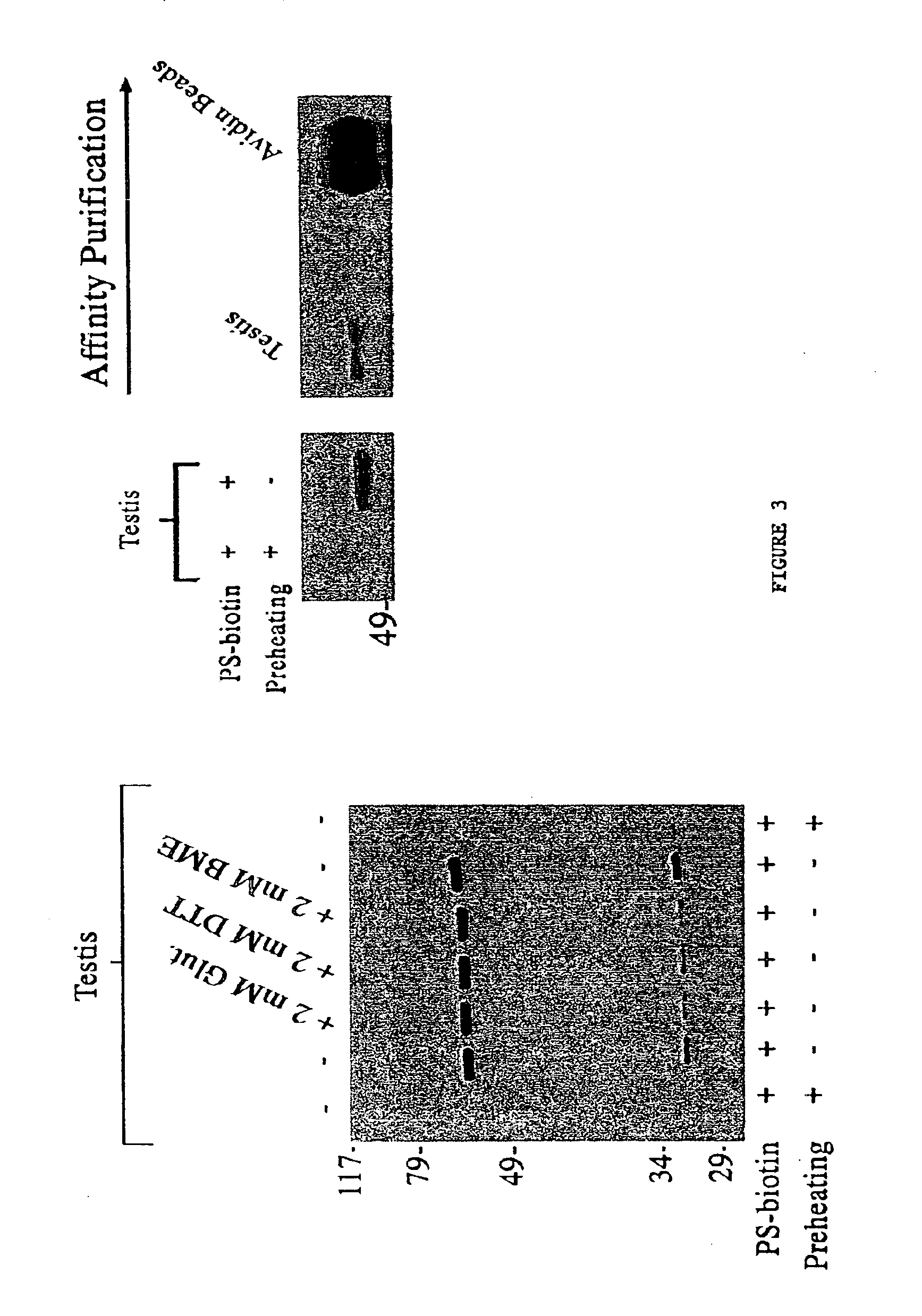
Brain soluble extracts were run over a Q sepharose column using an ÄKTA FPLC (Amersham Pharmacia Biotech) and eluted with a linear gradient of 0-500 mM NaCl. Samples of the elution fractions (10×2.5 mL fractions) were labeled with FP-biotin as described above, and those fractions containing the 75 kDa and 85 kDa labeled proteins were pooled and passed over a Mono-Q sepharose column. Proteins were eluted from the Mono-Q column with a linear gradient of 200-500 mM NaCl and those elution fractions enriched in the two labeled proteins were then run on SDS-PAGE and transferred to polyvinylidine difluoride (PVDF) membranes by electroblotting. Regions of the PVDF membranes containing the 75 and 85 kDa FP-biotin reactive proteins were excised, digested with trypsin, and the resulting peptides analyzed by matrix-assisted laser desorption ionization (MALDI) and MALDI-post-source decay time-of-flight mass spectrometry (Chaurand, et al. (...


PUM

PropertyMeasurementUnit
Fractionaaaaaaaaaa
Fractionaaaaaaaaaa
Fractionaaaaaaaaaa

Abstract

The present invention provides methods for analyzing proteomes, as cells or lysates. The analysis is based on the use of probes that have specificity to the active form of proteins, particularly enzymes and receptors. The probes can be identified in different ways. In accordance with the present invention, a method is provided for generating and screening compound libraries that are used for the identification of lead molecules, and for the parallel identification of their biological targets. By appending specific functionalities and / or groups to one or more binding moieties, the reactive functionalities gain binding affinity and specificity for particular proteins and classes of proteins. Such libraries of candidate compounds, referred to herein as activity-based probes, or ABPs, are used to screen for one or more desired biological activities or target proteins.

Description

FIELD OF THE INVENTIONThe field of this invention is analyzing portions of a proteome.BACKGROUND OF THE INVENTIONDetermination of the genomic sequence of higher organisms, including humans, is now an attainable goal. However, this analysis represents only one aspect of the information encoded by the genome. Genes are expressed in an ordered and timely manner, and are also exhibit a precise spatial and temporal expression pattern. Consequently, knowing the sequence of the genome is insufficient to explain biology and to understand disease. More significantly, genes are transcribed to messenger RNA, which is then translated to protein. It is the protein, or gene product, that exhibits activity, and carries out the work of the cell. With the post-genome era rapidly approaching, new strategies for the analysis of proteins are being developed. Most conventional approaches focus on recording variations in protein level. These approaches are commonly referred to as “proteomics”. In general...

Claims


Application Information

Patent Timeline
IPC IPC(8): C12Q1/37C12Q1/26C12Q1/32G01N33/535G01N33/557G01N33/68G06F19/00G01N33/50C07C309/66C07C309/73C07D495/04C07K14/00C12Q1/25C12Q1/34C40B20/04C40B30/04C40B40/02C40B40/04C40B50/06G01N33/15G16B20/30
CPCC12Q1/26C12Q1/32C12Q1/37C40B30/04C40B40/04G01N33/535G01N33/6842G01N33/6845G06F19/18G01N33/557Y10T436/16G01N2500/20Y10T436/18G16B20/00G16B20/30
Inventor CRAVATT, BENJAMIN F.SORENSEN, ERIKPATRICELLI, MATTHEW P.LOVATO, MARTHAADAM, GREGORY
Owner THE SCRIPPS RES INST
Learn More