Methods for spatial analysis using rna-templated ligation
a technology of rna-templated ligation and spatial analysis, which is applied in the direction of microbiological testing/measurement, biochemistry apparatus and processes, etc., can solve the problems of increasing the cost of an experiment, providing a lot of information, and not being human-friendly to formamide, so as to achieve the effect of less affected by rna degradation
- Summary
- Abstract
- Description
- Claims
- Application Information
AI Technical Summary
Benefits of technology
Problems solved by technology
Method used
Image
Examples
example 1
ene Expression Analysis of FFPE-Fixed Samples Using RNA-Templated Ligation
[0364]Others have demonstrated in situ ligation. See Credle et al., Nucleic Acids Research, Volume 45, Issue 14, 21 Aug. 2017, Page e128, https: / / doi.org / 10.1093 / nar / gkx471 (2017). However, the previous approaches have utilized hybridization using poly(A) tails which led to off-target binding. Here, the poly(A) tail on one probe oligonucleotide was switched to another sequence.
[0365]As an overview, a non-limiting example of RNA-templated ligation on an FFPE-fixed sample was performed as described in FIG. 13. FFPE-fixed samples were deparaffinized, stained (e.g., H&E stain), and imaged 1301. Samples were destained (e.g., using HCl) and decrosslinked 1302. Following decrosslinking, samples treated with pre-hybridization buffer (e.g., hybridization buffer without the first and second probes), probes were added to the sample, probes hybridized, and samples were washed 1303. Ligase was added to the samples to ligat...
example 2
ene Expression Analysis of Triple Positive Breast Cancer (TPBC) Using RNA Templated Ligation (RTL)
[0380]This example demonstrates that RTL can be performed on a sample in order to identify analyte abundance and spatial location in an unbiased manner.
[0381]A two-year old triple positive (“TPBC,” HER2, estrogen receptor, and progesterone receptor-positive) breast cancer sample preserved by FFPE processing was examined for analyte abundance and spatial location. The TPBC samples were queried with DNA probes via RNA-templated ligation methods. Before the ligation step, the TPBC tissue samples were deparaffinized and stained per established protocols. For example, FFPE TPBC tissue samples were prewarmed in a water bath (40° C.), sectioned (10 μm), dried at 42° C. for several hours and placed in a desiccator at room temperature overnight. The dry, sectioned tissues were deparaffinized by baking at 60° C., moved through a series of xylene and EtOH washes, rinsed in water several times. Fol...
embodiment b
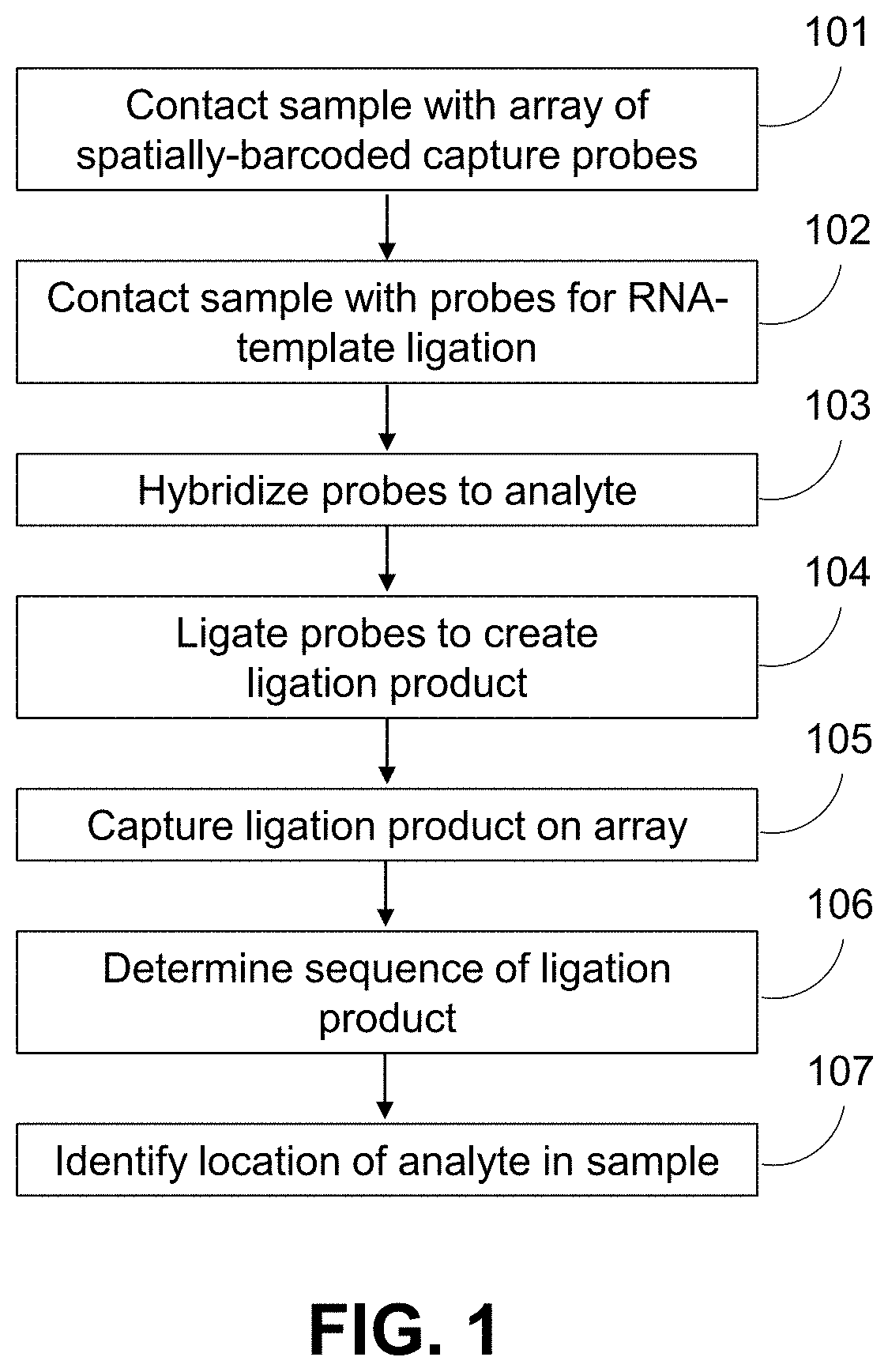
[0457]Embodiment B1. A method for identifying a location of an analyte in a biological sample, the method comprising:
[0458](a) contacting the biological sample with a substrate comprising a plurality of capture probes, wherein a capture probe of the plurality of capture probes comprises a capture domain and a spatial barcode;
[0459](b) contacting the biological sample with a first probe and a second probe, wherein a portion of the first probe and a portion of the second probe are substantially complementary to adjacent sequences of the analyte,
[0460]wherein the first probe comprises a sequence that is substantially complementary to a first target sequence of the analyte,
[0461]wherein the second probe comprises:[0462](i) a first sequence that is substantially complementary to a second target sequence of the analyte;[0463](ii) a linker sequence;[0464](iii) a second sequence that is substantially complementary to a third target sequence of the analyte; and[0465](iv) a capture probe capt...
PUM
| Property | Measurement | Unit |
|---|---|---|
| temperature | aaaaa | aaaaa |
| temperature | aaaaa | aaaaa |
| temperature | aaaaa | aaaaa |
Abstract
Description
Claims
Application Information
 - R&D
- Intellectual Property
- Life Sciences
- Materials
- Tech Scout
- Unparalleled Data Quality
- Higher Quality Content
- 60% Fewer Hallucinations
Browse by: Latest US Patents, China's latest patents, Technical Efficacy Thesaurus, Application Domain, Technology Topic, Popular Technical Reports.
© 2025 PatSnap. All rights reserved.Legal|Privacy policy|Modern Slavery Act Transparency Statement|Sitemap|About US| Contact US: help@patsnap.com


